. 1998 Sep;180(18):4967-73.
doi: 10.1128/JB.180.18.4967-4973.1998.

Characterization of dacC, which encodes a new low-molecular-weight penicillin-binding protein in Bacillus subtilis

Affiliations

Characterization of dacC, which encodes a new low-molecular-weight penicillin-binding protein in Bacillus subtilis

L B Pedersen et al. J Bacteriol. 1998 Sep.

Abstract

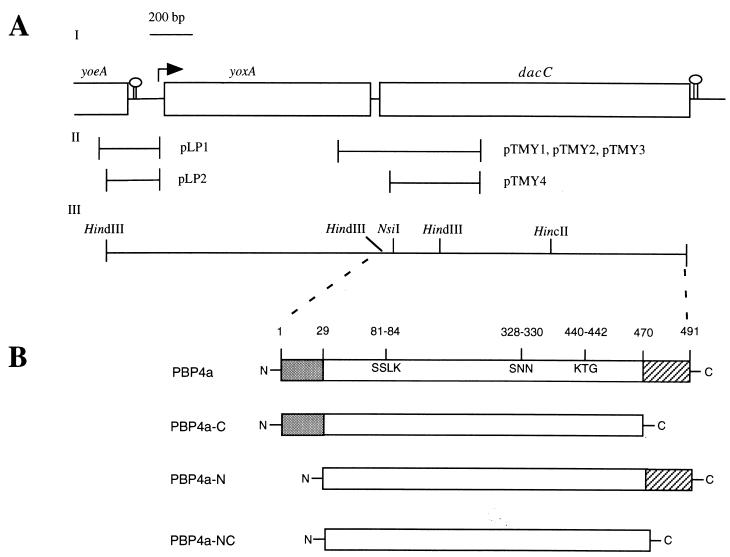
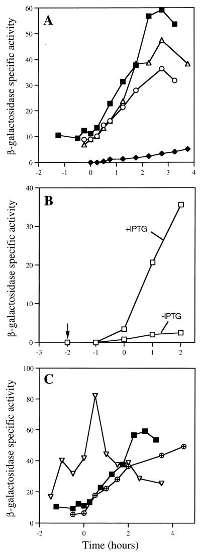
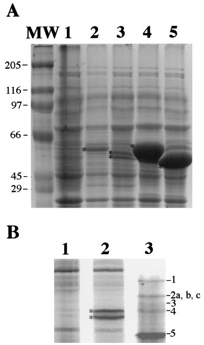
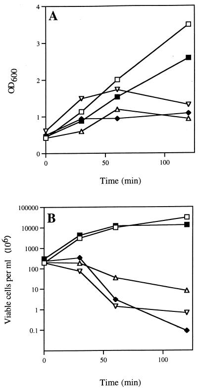
The pbp gene (renamed dacC), identified by the Bacillus subtilis genome sequencing project, encodes a putative 491-residue protein with sequence homology to low-molecular-weight penicillin-binding proteins. Use of a transcriptional dacC-lacZ fusion revealed that dacC expression (i) is initiated at the end of stationary phase; (ii) depends strongly on transcription factor sigmaH; and (iii) appears to be initiated from a promoter located immediately upstream of yoxA, a gene of unknown function located upstream of dacC on the B. subtilis chromosome. A B. subtilis dacC insertional mutant grew and sporulated identically to wild-type cells, and dacC and wild-type spores had the same heat resistance, cortex structure, and germination and outgrowth kinetics. Expression of dacC in Escherichia coli showed that this gene encodes an approximately 59-kDa membrane-associated penicillin-binding protein which is highly toxic when overexpressed.


Figures

FIG. 1
FIG. 1
Diagram of the dacC locus, and constructs and protein variants generated. (A) Map of the dacC locus. (I) Putative ORFs are indicated by open boxes, potential transcription terminators are shown as stem-loop structures, and the arrow depicts the predicted transcription initiation site and direction of transcription. (II) Fragments used in plasmid constructs for insertional mutagenesis and for generation of transcriptional dacC-lacZ fusions. (III) Map of selected restriction endonuclease cleavage sites. (B) Schematic depiction of PBP4a variants generated in this work. Amino acids 1 to 29 (gray) constitute a cleavable signal peptide as described in the text. The three regions that constitute the penicillin-binding site (81SSLK84, 328SNN330, and 440KTG442) were inferred by sequence alignment of PBP4a with PBP4 from Actinomadura strain R39 (14) and E. coli PBP4 (19) using GCG software (Wisconsin Package Version 9.1; Genetics Computer Group, Madison, Wis.). Amino acids 470 to 491 (hatched) were predicted by a computer analysis (DNA Strider 1.2) to form an amphipathic α-helix, potentially serving as a membrane anchor. Numbers refer to amino acids of the PBP4a primary sequence. The figure is not drawn to scale.
FIG. 2
FIG. 2
Transcriptional regulation of dacC. B. subtilis strains containing transcriptional dacC-lacZ fusions at the dacC locus were grown and sporulated at 37°C in 100 ml of 2× SG medium with no antibiotics (24), the OD600 values of the cultures were measured, and 1-ml samples were withdrawn for measurement of β-galactosidase activity as described in the text at the times indicated (t0 is defined as the end of exponential phase). Values on the y axis are fluorescence units per OD600 unit of the cultures. (A) β-Galactosidase activities in strains lacking sigma factors. Symbols and strains (relevant genotypes) are as follows: ■, PS2323 (wild type); ⧫, PS2459 (spo0H::Kanr); ○, PS2522 (ΔspoIIA::Spr); and ▵, PS2521 (ΔspoIIGB::Ermr). (B) β-Galactosidase activity in strain PS2629 (Pspac-spo0H) with or without IPTG (arrow indicates time of IPTG addition). (C) β-Galactosidase activities in transcription factor mutant strains. Symbols and strains (relevant genotypes) are as follows: ▿, PS2632 (spo0A::Ermr); ⊕, PS2630 (abrB::Tn917); and ■, PS2323 (wild type).
FIG. 3
FIG. 3
Expression of dacC variants and penicillin-binding activity of PBP4a. (A) Expression of dacC variants in E. coli. Recombinant E. coli strains were grown and induced, protein was solubilized as described in the text, and 7 μl of each sample was analyzed by SDS–10% PAGE and staining with Coomassie blue. Lanes, corresponding strains, and proteins they express (parentheses) are as follows: 1, PS2602 (vector alone); 2, PS2599 (PBP4a); 3, PS2690 (PBP4a-C); 4, PS2691 (PBP4a-N); and 5, PS2692 (PBP4a-NC). Lane MW contains molecular weight markers (molecular masses are in kilodaltons). Asterisks denote migration positions of the PBP4a variants. (B) Analysis of PBPs in membranes from induced E. coli strains PS2602 (lane 1; vector alone) and PS2599 (lane 2; PBP4a). Cells were grown and induced as for panel A for 90 min, 25 ml of culture was harvested by centrifugation, membranes were isolated from sonicated cells by centrifugation (100,000 × g, 1 h) and incubated for 30 min at 30°C with 100 μM FLU-C6-APA, proteins (∼10 μg) were analyzed by SDS–10% PAGE, and PBPs were visualized with a FluorimagerSI (Vistra). A lane containing labeled PBPs from vegetative B. subtilis cells of strain PS832 (32) is shown for comparison (lane 3; the PBPs corresponding to each band are indicated on the right). Asterisks denote position of the 59–60-kDa PBP4a doublet.
FIG. 4
FIG. 4
Effects of PBP4a variants on growth and viability of E. coli. Recombinant E. coli strains were grown and induced with IPTG as described in the text, and the OD600 (A) and viability (B) were measured after induction. The viability was measured by plating dilutions on 2× YT agar plates containing chloramphenicol (20 μg ml−1) and ampicillin (50 μg ml−1). Symbols, strains, and proteins they express (parentheses) are as follows: □, PS2602 (vector); ▿, PS2599 (PBP4a); ▵, PS2690 (PBP4a-C); ⧫, PS2691 (PBP4a-N); and ■, PS2692 (PBP4a-NC).

Similar articles

Cited by

References

    1. Adam M, Damblon C, Plaitin B, Christiaens L, Frère J-M. Chromogenic depsipeptide substrates for beta-lactamases and penicillin-sensitive DD-peptidases. Biochem J. 1990;270:525–529. - PMC - PubMed
    1. Anagnostopoulos C, Spizizen J. Requirements for transformation in Bacillus subtilis. J Bacteriol. 1961;81:741–746. - PMC - PubMed
    1. Antoniewski C, Savelli B, Stragier P. The spoIIJ gene, which regulates early developmental steps in Bacillus subtilis, belongs to a class of environmentally responsive genes. J Bacteriol. 1990;172:86–93. - PMC - PubMed
    1. Atrih A P Z, Allmaier G, Foster S. Structural analysis of Bacillus subtilis endospore peptidoglycan and its role during differentiation. J Bacteriol. 1996;178:6173–6183. - PMC - PubMed
    1. Blumberg P M, Strominger J L. Inactivation of D-alanine carboxypeptidase by penicillins and cephalosporins is not lethal in Bacillus subtilis. Proc Natl Acad Sci USA. 1971;68:2814–2817. - PMC - PubMed

Publication types

LinkOut - more resources