

Characterization of and functional antigen presentation by central nervous system mononuclear cells from mice infected with Theiler's murine encephalomyelitis virus
- PMID: 9733812
- PMCID: PMC110086
- DOI: 10.1128/JVI.72.10.7762-7771.1998
Characterization of and functional antigen presentation by central nervous system mononuclear cells from mice infected with Theiler's murine encephalomyelitis virus
Abstract
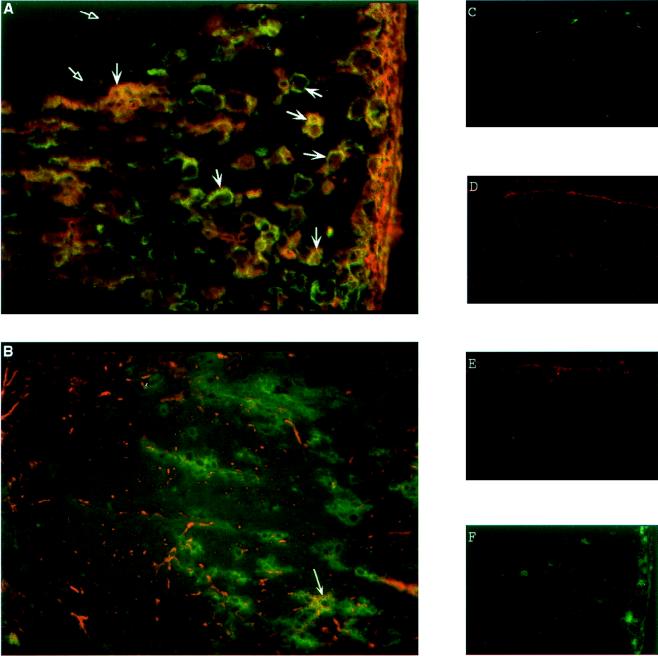
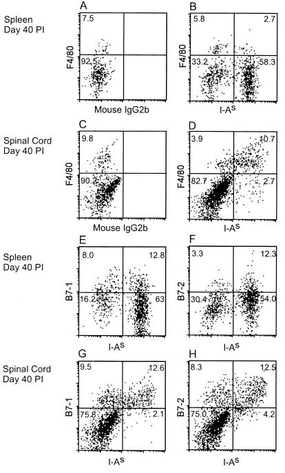
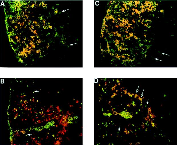
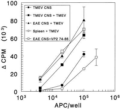
We examined the phenotype and function of cells infiltrating the central nervous system (CNS) of mice persistently infected with Theiler's murine encephalomyelitis virus (TMEV) for evidence that viral antigens are presented to T cells within the CNS. Expression of major histocompatibility complex (MHC) class II in the spinal cords of mice infected with TMEV was found predominantly on macrophages in demyelinating lesions. The distribution of I-As staining overlapped that of the macrophage marker sialoadhesin in frozen sections and coincided with that of another macrophage/microglial cell marker, F4/80, by flow cytometry. In contrast, astrocytes, identified by staining with glial fibrillary acidic protein, rarely expressed detectable MHC class II, although fibrillary gliosis associated with the CNS damage was clearly seen. The costimulatory molecules B7-1 and B7-2 were expressed on the surface of most MHC class II-positive cells in the CNS, at levels exceeding those found in the spleens of the infected mice. Immunohistochemistry revealed that B7-1 and B7-2 colocalized on large F4/80(+) macrophages/microglia in the spinal cord lesions. In contrast, CD4(+) T cells in the lesions expressed mainly B7-2, which was found primarily on blastoid CD4(+) T cells located toward the periphery of the lesions. Most interestingly, plastic-adherent cells freshly isolated from the spinal cords of TMEV-infected mice were able to process and present TMEV and horse myoglobin to antigen-specific T-cell lines. Furthermore, these cells were able to activate a TMEV epitope-specific T-cell line in the absence of added antigen, providing conclusive evidence for the endogenous processing and presentation of virus epitopes within the CNS of persistently infected SJL/J mice.
Figures

Similar articles
-
Temporal development of autoreactive Th1 responses and endogenous presentation of self myelin epitopes by central nervous system-resident APCs in Theiler's virus-infected mice.J Immunol. 2000 Nov 1;165(9):5304-14. doi: 10.4049/jimmunol.165.9.5304. J Immunol. 2000. PMID: 11046065
-
CNS expression of B7-H1 regulates pro-inflammatory cytokine production and alters severity of Theiler's virus-induced demyelinating disease.PLoS One. 2011 Apr 8;6(4):e18548. doi: 10.1371/journal.pone.0018548. PLoS One. 2011. PMID: 21494618 Free PMC article.
-
Endogenous presentation of self myelin epitopes by CNS-resident APCs in Theiler's virus-infected mice.J Clin Invest. 1999 Sep;104(5):599-610. doi: 10.1172/JCI7292. J Clin Invest. 1999. PMID: 10487774 Free PMC article.
-
Theiler's virus-mediated autoimmunity: local presentation of CNS antigens and epitope spreading.Ann N Y Acad Sci. 2002 Apr;958:26-38. Ann N Y Acad Sci. 2002. PMID: 12021081 Review.
-
The functional significance of epitope spreading and its regulation by co-stimulatory molecules.Immunol Rev. 1998 Aug;164:63-72. doi: 10.1111/j.1600-065x.1998.tb01208.x. Immunol Rev. 1998. PMID: 9795764 Review.
Cited by
-
Disproportionate recruitment of CD8+ T cells into the central nervous system by professional antigen-presenting cells.Am J Pathol. 1999 Feb;154(2):481-94. doi: 10.1016/S0002-9440(10)65294-7. Am J Pathol. 1999. PMID: 10027406 Free PMC article.
-
Regulation and function of class II major histocompatibility complex, CD40, and B7 expression in macrophages and microglia: Implications in neurological diseases.J Neurovirol. 2002 Dec;8(6):496-512. doi: 10.1080/13550280290100941. J Neurovirol. 2002. PMID: 12476345 Review.
-
Role of peripheral immune response in microglia activation and regulation of brain chemokine and proinflammatory cytokine responses induced during VSV encephalitis.J Neuroimmunol. 2014 Feb 15;267(1-2):50-60. doi: 10.1016/j.jneuroim.2013.12.002. Epub 2013 Dec 11. J Neuroimmunol. 2014. PMID: 24369299 Free PMC article.
-
Machine learning approach identifies new pathways associated with demyelination in a viral model of multiple sclerosis.J Cell Mol Med. 2010 Jan;14(1-2):434-48. doi: 10.1111/j.1582-4934.2008.00646.x. Epub 2009 Jan 14. J Cell Mol Med. 2010. PMID: 19183246 Free PMC article.
-
Selection of and evasion from cytotoxic T cell responses in the central nervous system.Adv Virus Res. 2001;56:219-42. doi: 10.1016/s0065-3527(01)56029-7. Adv Virus Res. 2001. PMID: 11450301 Free PMC article. Review.
References
-
- Clatch R J, Melvold R W, Dal Canto M C, Miller S D, Lipton H L. The Theiler’s murine encephalomyelitis virus (TMEV) model for multiple sclerosis shows a strong influence of the murine equivalents of HLA-A, B, and C. J Neuroimmunol. 1987;15:121–135. - PubMed
-
- Clatch R J, Miller S D, Metzner R, Dal Canto M C, Lipton H L. Monocytes/macrophages isolated from the mouse central nervous system contain infectious Theiler’s murine encephalomyelitis virus (TMEV) Virology. 1990;176:244–254. - PubMed
Publication types
MeSH terms
Substances
Grants and funding
LinkOut - more resources
Full Text Sources
Research Materials
