. 1998 Oct;72(10):8089-97.
doi: 10.1128/JVI.72.10.8089-8097.1998.

Avian hepatitis B virus infection is initiated by the interaction of a distinct pre-S subdomain with the cellular receptor gp180

Affiliations

Avian hepatitis B virus infection is initiated by the interaction of a distinct pre-S subdomain with the cellular receptor gp180

S Urban et al. J Virol. 1998 Oct.

Abstract

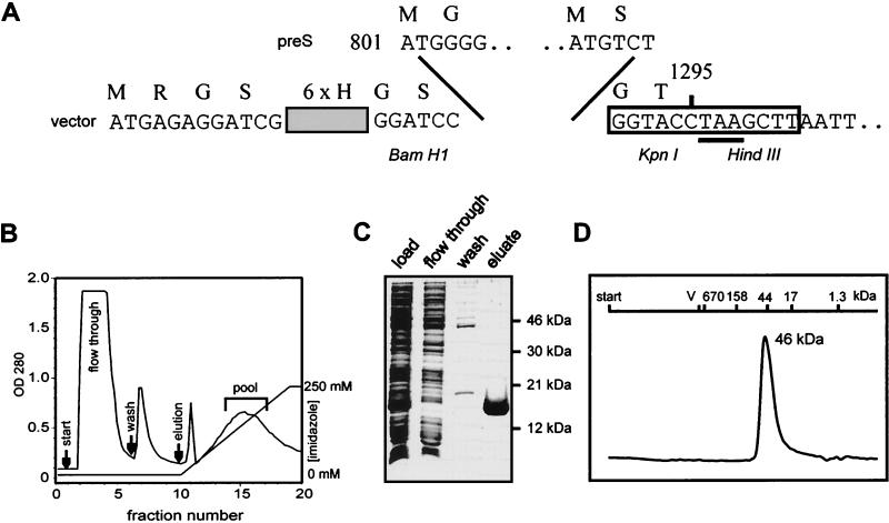
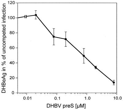
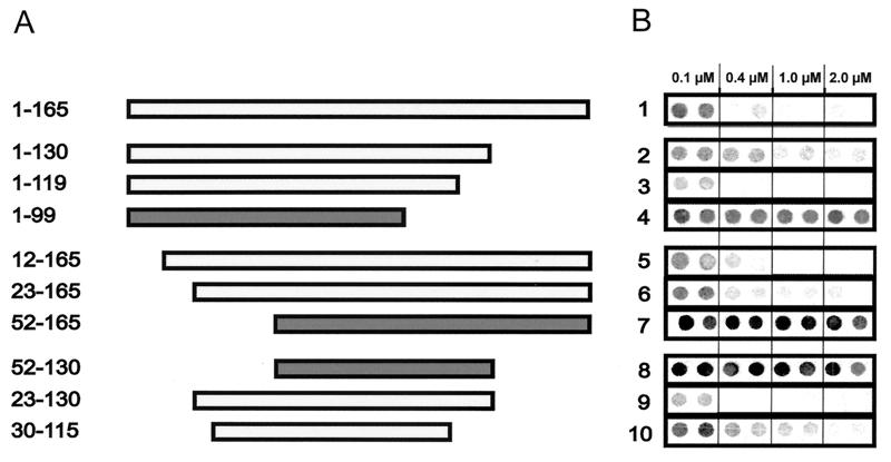
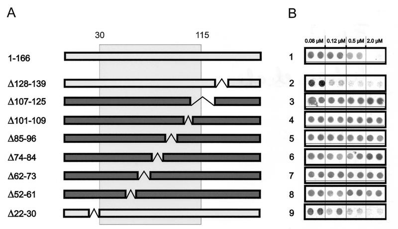
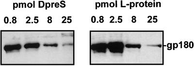
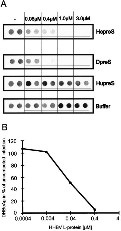
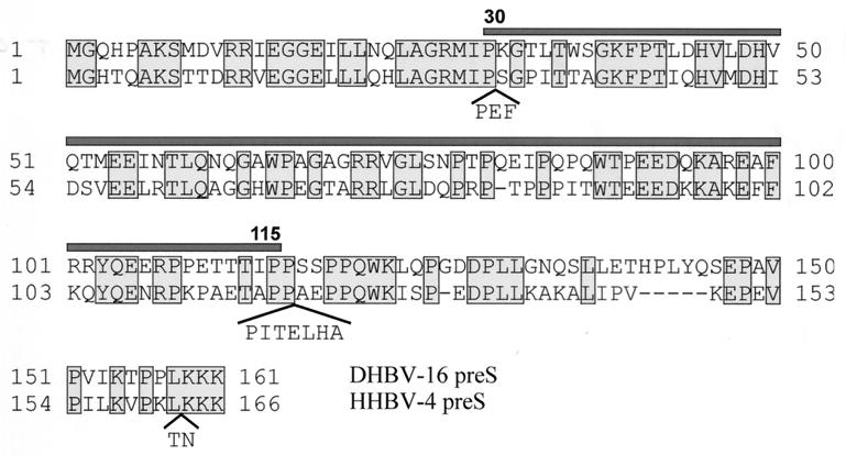
Functionally relevant hepadnavirus-cell surface interactions were investigated with the duck hepatitis B virus (DHBV) animal model by using an in vitro infection competition assay. Recombinant DHBV pre-S polypeptides, produced in Escherichia coli, were shown to inhibit DHBV infection in a dose-dependent manner, indicating that monomeric pre-S chains were capable of interfering with virus-receptor interaction. Particle-associated pre-S was, however, 30-fold more active, suggesting that cooperative interactions enhance particle binding. An 85-amino-acid pre-S sequence, spanning about half of the DHBV pre-S chain, was characterized by deletion analysis as essential for maximal inhibition. Pre-S polypeptides from heron hepatitis B virus (HHBV) competed DHBV infection equally well despite a 50% difference in amino acid sequence and a much-reduced infectivity of HHBV for duck hepatocytes. These observations are taken to indicate (i) that the functionality of the DHBV pre-S subdomain, which interacts with the cellular receptor, is determined predominantly by a defined three-dimensional structure rather than by primary sequence elements; (ii) that cellular uptake of hepadnaviruses is a multistep process involving more than a single cellular receptor component; and (iii) that gp180, a cellular receptor candidate unable to discriminate between DHBV and HHBV, is a common component of the cellular receptor complex for avian hepadnaviruses.


Figures

FIG. 1
FIG. 1
Expression of pre-S-coding sequences in E. coli and purification of recombinant proteins. (A) Pre-S-coding sequences were inserted into the prokaryotic expression vector pQE 8 (Qiagen) modified by a KpnI/HindIII linker (boxed sequence) as described in Materials and Methods. The construct encoding full-length pre-S and the first 4 aa from the S region is shown. Numbers 801 and 1295 indicate the first and the last nucleotides of the subcloned fragment, which is followed by a stop codon (underlined). The six amino-terminal histidine residues and six additional vector-encoded amino acids are shown in one-letter code above the nucleotide sequence. (B) Affinity chromatography with an Ni2+-nitrilotriacetic acid column. Hexahistidine pre-S fusion proteins were purified from total cellular lysates of recombinant E. coli as described in Materials and Methods and shown here for the DHBV pre-S fragment from aa 1 to 119. Elution was performed with an imidazole gradient from 0 to 250 mM as indicated. Pooled fractions are marked by a bracket. OD280, optical density at 280 nm. (C) Silver-stained SDS gel of fractions eluted from the affinity column shown in panel B. Molecular masses of marker proteins are indicated on the right. (D) Elution profile of renatured DHBV pre-S on a Superdex 200 column (Pharmacia). V0 and the elution volumes of thyroglobulin (670 kDa), gamma globulin (158 kDa), ovalbumin (44 kDa), myoglobin (17 kDa), and vitamin B12 (1.3 kDa), used for calibration, are indicated at the top.
FIG. 2
FIG. 2
Competition of DHBV infection by recombinant DHBV pre-S polypeptide. PDHs (8 × 10⁵) were infected with 4 × 10⁷ DHBV particles in the presence of increasing concentrations of E. coli-derived DHBV pre-S protein (subtype 16). Virus replication between days 5 and 9 postinfection was determined by immuno-dot blot analysis of DHBeAg secreted into the culture medium and is presented as a percentage of the value for an uncompeted control infection (for details, see Materials and Methods). Each point represents the average of three independent experiments; bars indicate the standard deviations.
FIG. 3
FIG. 3
Competition of DHBV infection by terminally deleted DHBV pre-S polypeptides. PDHs (8 × 10⁵) were infected with 4 × 10⁷ DNA-containing particles in the presence of increasing concentrations of either full-length or terminally deleted recombinant DHBV pre-S polypeptides. (A) Schematic drawing of the amino- and carboxy-terminal deletion mutants tested. Numbers on the left indicate the limits of the polypeptides, which are depicted as bars. Note that the entire pre-S sequence covers aa 1 to 161 (Fig. 1A). Bars in light gray correspond to polypeptides that behave in infection inhibition like full-length polypeptide (aa 1 to 165); bars in dark gray represent polypeptide mutants that lost activity. (B) DHBeAg dot blot analysis of culture medium from PDHs collected between days 5 and 9 postinfection in the presence of the pre-S polypeptides depicted in panel A. Polypeptide concentrations are indicated at the top. DHBeAg was quantified as described in Materials and Methods. Each dot corresponds to a single well of a 12-well culture plate. Two independent experiments for each concentration are shown. Apparent differences in competition activity relative to the reference in row 1 (visible in rows 2 and 10) were preparation dependent and could not be reproduced in independent experiments using another polypeptide preparation.
FIG. 4
FIG. 4
Competition of DHBV infection of PDHs by internally deleted pre-S polypeptides. (A) Schematic drawing of the mutant polypeptides (DHBV pre-S subtype 26). Deletions are shown by gaps within the bars. Bars in light gray indicate protein mutants that behave like full-length pre-S; bars in dark gray represent protein mutants that lost their ability to compete infection. The minimal competitor region (aa 30 to 115) as determined by analysis of terminally deleted DHBV pre-S polypeptides is indicated as a box. Note that DHBV pre-S subtype 26 encodes an additional amino acid at position 146. The full-length protein therefore contains 166 aa. (B) Immuno-dot blot analysis of culture medium from PDHs in the presence of different internal pre-S mutants (rows 2 to 9) compared with full-length DHBV pre-S (row 1). DHBeAg was quantified as described in Materials and Methods. Two independent experiments for each concentration are shown.
FIG. 5
FIG. 5
Recombinant DHBV pre-S polypeptide binds gp180 with affinities comparable to those of particle-derived DHBV L protein. Western blots detecting gp180, bound to immobilized DHBV pre-S polypeptide in the presence of increasing concentrations of competing free DHBV pre-S polypeptide (DpreS) or DHBV L protein derived from serum of a DHBV-infected duckling (L-protein), are shown. Numbers above the lanes indicate the amounts of free competitor present during binding of gp180 to immobilized DHBV pre-S. The gp180-specific band is indicated at the right.
FIG. 6
FIG. 6
Competition of DHBV infection with heterologous pre-S proteins. Infection competition was carried out as described in the legends to Fig. 2 and 3. (A) Immuno-dot blot analysis of culture medium from PDHs between days 5 and 9 postinfection with DHBV in the presence of HHBV pre-S polypeptide (HepreS), DHBV pre-S polypeptide (DpreS), HBV pre-S polypeptide (HupreS), or buffer. Polypeptide concentrations are indicated at the top. For each concentration, two independent measurements are shown. (B) Competition of DHBV infection by SVPs from HHBV. Amounts of secreted DHBeAg are presented as a percentage of the value for an uncompeted control infection.
FIG. 7
FIG. 7
Comparison of the amino acid sequences of the pre-S domains of DHBV (subtype 16, upper row) and HHBV (subtype 4, lower row). Amino acid positions are indicated by numbers beside the rows. Conserved amino acids are boxed. Insertions within the HHBV pre-S sequence are shown in brackets. The minimal competitor region representing the receptor binding domain (aa 30 to 115) is indicated by the bar at the top of the DHBV pre-S sequence.

References

    1. Breiner K M, Urban S, Schaller H. Carboxypeptidase D (gp180), a Golgi-resident protein, functions in the attachment and entry of avian hepatitis B viruses. J Virol. 1998;72:8094–8104. - PMC - PubMed
    1. DeMeyer S, Gong J Z, Suwandhi W, van Pelt J, Soumillon A, Yap S H. Organ and species specificity of hepatitis B virus (HBV) infection: a review of literature with a special reference to preferential attachment of HBV to human hepatocytes. J Viral Hepat. 1997;4:145–153. - PubMed
    1. Deng H, Liu R, Ellmeier W, Choe S, Unutmaz D, Burkhart M, Di M P, Marmon S, Sutton R E, Hill C M, Davis C B, Peiper S C, Schall T J, Littman D R, Landau N R. Identification of a major co-receptor for primary isolates of HIV-1. Nature. 1996;381:661–666. - PubMed
    1. Fehler, F., S. Seitz, and H. Schaller. Unpublished data.
    1. Feng Y, Broder C C, Kennedy P E, Berger E A. HIV-1 entry cofactor: functional cDNA cloning of a seven-transmembrane, G protein-coupled receptor. Science. 1996;272:872–877. - PubMed

Publication types

MeSH terms