. 1998 Oct;72(10):8150-7.
doi: 10.1128/JVI.72.10.8150-8157.1998.

Development of a self-inactivating lentivirus vector

Affiliations

Development of a self-inactivating lentivirus vector

H Miyoshi et al. J Virol. 1998 Oct.

Abstract

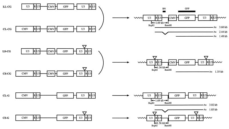
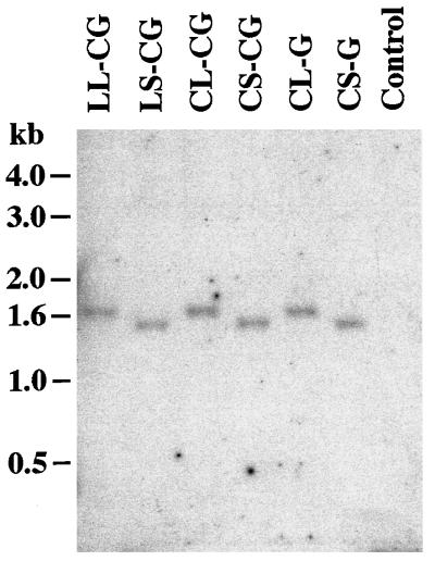
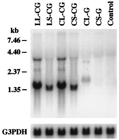
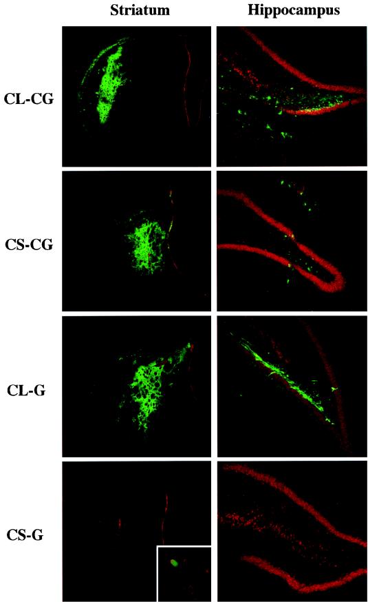
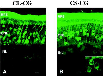
We have constructed a new series of lentivirus vectors based on human immunodeficiency virus type 1 (HIV-1) that can transduce nondividing cells. The U3 region of the 5' long terminal repeat (LTR) in vector constructs was replaced with the cytomegalovirus (CMV) promoter, resulting in Tat-independent transcription but still maintaining high levels of expression. A self-inactivating (SIN) vector was constructed by deleting 133 bp in the U3 region of the 3' LTR, including the TATA box and binding sites for transcription factors Sp1 and NF-kappaB. The deletion is transferred to the 5' LTR after reverse transcription and integration in infected cells, resulting in the transcriptional inactivation of the LTR in the proviruses. SIN viruses can be generated with no significant decreases in titer. Injection of viruses into the rat brain showed that a SIN vector containing the green fluorescent protein gene under the control of the internal CMV promoter transduced neurons as efficiently as a wild-type vector. Interestingly, a wild-type vector without an internal promoter also successfully transduced neurons in the brain, indicating that the HIV-1 LTR promoter is transcriptionally active in neurons even in the absence of Tat. Furthermore, injection of viruses into the subretinal space of the rat eye showed that wild-type vector transduced predominantly retinal pigment epithelium and photoreceptor cells, while SIN vector was able to transduce other types of retinal cells, including bipolar, Müller, horizontal, and amacrine cells. This finding suggests that the HIV-1 LTR can negatively influence the internal CMV promoter in some cell types. SIN HIV vectors should be safer for gene therapy, and they also have broader applicability as a means of high-level gene transfer and expression in nondividing cells.


Figures

FIG. 1
FIG. 1
Structures of HIV vector constructs and corresponding proviruses. (Left) HIV vector constructs. Each vector construct is cotransfected with the packaging and VSV-G expression constructs into 293T cells. Viral transcription initiates at the U3/R border in the 5′ LTR and terminates at the R/U5 border in the 3′ LTR. The viral RNA is packaged into virions. Virus is harvested and used to infect target cells. Triangles represent deleted U3 region. (Right) Structures of integrated proviruses. In infected cells, the U3 region of the 3′ LTR is used as a template for the synthesis of the U3 region in both LTRs during the process of reverse transcription of the viral RNA into double-stranded DNA. As a result, the U3 region of the 3′ LTR is duplicated and transferred to the 5′ LTR in the integrated provirus. The BspEI-BamHI restriction fragments expected in Southern blot analysis (Fig. 2) and the RNA transcripts expected in Northern blot analysis (Fig. 3) are shown below each proviral structure with their sizes. The locations of the BN and GFP probes used in Southern blot and Northern blot analyses are also indicated.
FIG. 2
FIG. 2
Southern blot analysis of the integrated proviral structure. Genomic DNA isolated from HeLa-CD4-LTR-β-gal cells infected with HIV vectors was digested with BspEI and BamHI. The blot was hybridized with the BN probe. The vector used for infection is indicated above each lane. Control, uninfected HeLa-CD4-LTR-β-gal cells. The expected sizes of fragments are shown in Fig. 1. Size markers are indicated on the left.
FIG. 3
FIG. 3
Northern blot analysis of the proviral expression. Total cellular RNA was isolated from HeLa-CD4-LTR-β-gal cells infected with HIV vectors. The blot was hybridized with the GFP probe and rehybridized with the human glyceraldehyde-3-phosphate dehydrogenase (G3PDH) probe (Clontech). The vector used for infection is indicated above each lane. Control, uninfected HeLa-CD4-LTR-β-gal cells. The expected size of each transcript is shown in Fig. 1. Size markers are indicated on the left.
FIG. 4
FIG. 4
Expression of GFP in the striatum and hippocampus of adult rat brains 6 weeks after injection of HIV vectors. Sections were counterstained with propidium iodide. The vectors used for injection are indicated on the left. Inset, higher magnification of a GFP-positive cell.
FIG. 5
FIG. 5
Expression of GFP in the retina of rat pups 12 weeks after injection of the CL-CG vector (A) and the CS-CG vector (B). Inset, higher magnification of GFP-positive inner nuclear layer (INL) cells. Scale bars, 10 μm. RPE, retinal pigment epithelium; ONL, outer nuclear layer.

References

    1. Armentano D, Yu S-F, Kantoff P W, Ruden T V, Anderson W F, Gilboa E. Effect of internal viral sequences on the utility of retroviral vectors. J Virol. 1987;61:1647–1650. - PMC - PubMed
    1. Berkhout B, Gatignol A, Rabson A B, Jeang K-T. TAR-independent activation of the HIV-1 LTR: evidence that tat requires specific regions of the promoter. Cell. 1990;62:757–767. - PubMed
    1. Berkhout B, Jeang K-T. Functional roles for the TATA promoter and enhancers in basal and tat-induced expression of the human immunodeficiency virus type 1 long terminal repeat. J Virol. 1992;66:139–149. - PMC - PubMed
    1. Blömer U, Naldini L, Kafri T, Trono D, Verma I M, Gage F H. Highly efficient and sustained gene transfer in adult neurons with a lentivirus vector. J Virol. 1997;71:6641–6649. - PMC - PubMed
    1. Bowtell D D L, Cory S, Johnson G R, Gonda T J. Comparison of expression in hemopoietic cells by retroviral vectors carrying two genes. J Virol. 1988;62:2464–2473. - PMC - PubMed

Publication types