. 1998 Oct;72(10):8198-204.
doi: 10.1128/JVI.72.10.8198-8204.1998.

Measles virus fusion protein is palmitoylated on transmembrane-intracytoplasmic cysteine residues which participate in cell fusion

Affiliations

Measles virus fusion protein is palmitoylated on transmembrane-intracytoplasmic cysteine residues which participate in cell fusion

M Caballero et al. J Virol. 1998 Oct.

Abstract

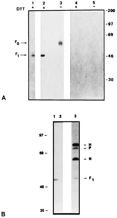
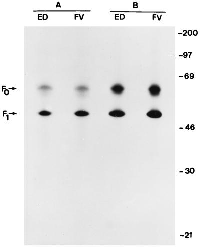
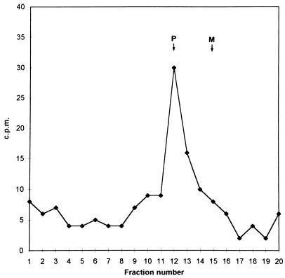
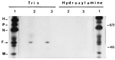
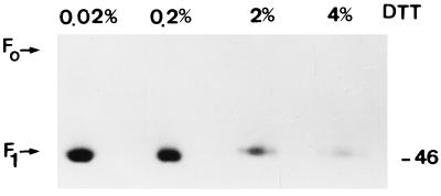
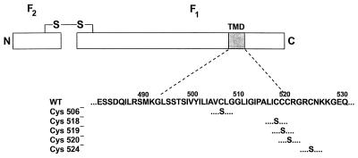
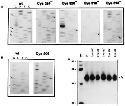
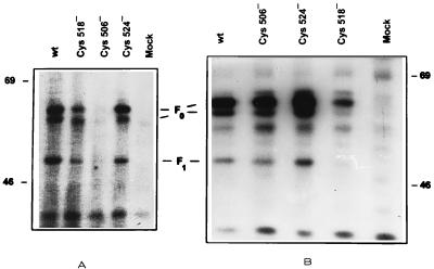
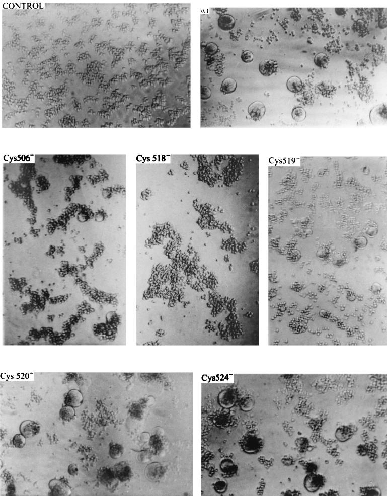
[3H]palmitic acid was metabolically incorporated into the viral fusion protein (F) of Edmonston or freshly isolated measles virus (MV) during infection of human lymphoid or Vero cells. The uncleaved precursor F0 and the F1 subunit from infected cells and extracellular virus were both labeled, indicating that palmitoylation can take place prior to F0 cleavage and that palmitoylated F protein was incorporated into virus particles. [3H]palmitic acid was released from F protein upon hydroxylamine or dithiothreitol treatment, indicating a thioester linkage. In cells transfected with the cloned MV F gene, in which the cysteines located in the intracytoplasmic and transmembrane domains (Cys 506, 518, 519, 520, and 524) were replaced by serine, a major reduction of [3H]palmitic acid incorporation was observed for F mutated at Cys 506 and, to a lesser extent, at Cys 518 and Cys 524. We also observed incorporation of [3H]palmitic acid in the F1 subunit of canine distemper virus F protein. Cell fusion induced by cotransfection of cells with MV F and H (hemagglutinin) genes was significantly reduced after replacement of Cys 506 or Cys 519 with serine in the MV F gene. Transfection with the F gene with a mutation for Cys 518 abolished cell fusion, although less mutant protein was detected on the cell surface. These results suggest that the F protein transmembrane domain cysteines 506 and 518 participate in structures involved in cell fusion, possibly mediated by palmitoylation.


Figures

FIG. 1
FIG. 1
(A) PAGE of [3H]palmitic acid (lanes 1 to 3)- and [3H]myristic acid (lanes 4 and 5)-labeled polypeptides in Edmonston MV-infected MOLT4 cells. Extracellular (lane 1) and intracellular (lanes 2 to 5) viral proteins were labeled for 15 h, immunoprecipitated with anti-MV guinea pig serum, and analyzed under reducing (+) or nonreducing (−) conditions. A 25-day exposure of the fluorogram is shown. Molecular masses (in kilodaltons) are on the right. (B) Incorporation of [3H]palmitic acid into F protein of CDV. [3H]palmitic acid-labeled intracellular polypeptides in uninfected (lane 2) and CDV-infected (lane 1) Vero cells and TRAN35S-labeled proteins from CDV-infected Vero cells (lane 3) are shown. CDV proteins were immunoprecipitated with anti-MV guinea pig serum and analyzed in a SDS–10% polyacrylamide slab gel. A 30-day exposure of the fluorogram is shown. Molecular masses (in kilodaltons) are on the left.
FIG. 2
FIG. 2
Incorporation of [3H]palmitic acid into F protein of Edmonston strain and wild-type isolate FV. [3H]palmitic acid-labeled intracellular polypeptides in Edmonston (ED) and primary isolate Ma93F (FV) virus-infected Dakiki cells (2 × 10⁶) were immunoprecipitated with the anti-F MAb Ost-2 (A), and the supernatant was immunoprecipitated with the anti-MV guinea-pig serum (B). A 20-day exposure of the fluorogram is shown. Molecular masses (in kilodaltons) are on the right.
FIG. 3
FIG. 3
Identification of fatty acid removed from F protein as palmitate. Thin-layer chromatography of fatty acids from hydrolyzed [3H]palmitic acid-labeled F protein from MV Edmonston-infected MOLT4 cells. Markers of palmitic (P) and myristic (M) acids were detected under UV light after rhodamine G impregnation, and radioactivity was estimated in a beta radiation counter.
FIG. 4
FIG. 4
Sensitivity of MV acylated proteins to hydroxylamine. Extracts from MV Edmonston-infected MOLT4 cells labeled with TRAN35S-label (1) or [3H]palmitate (2 and 3) were immunoprecipitated with anti-MV antibodies; the immunocomplexes were treated with 1 M Tris-HCl (pH 8) or 1 M hydroxylamine (pH 8) at room temperature for 3 h and analyzed by SDS-PAGE and fluorography. Molecular masses (in kilodaltons) are on the right.
FIG. 5
FIG. 5
Sensitivity of acylated F protein to DTT. Fusion protein immunocomplexes from [3H]palmitic acid-labeled MV Edmonston-infected MOLT4 cells were treated with increasing concentrations of DTT. Samples were heated to 95°C for 5 min and subjected to SDS-PAGE and fluorography. The molecular mass (in kilodaltons) is on the right.
FIG. 6
FIG. 6
Schematic diagram of mutations introduced at the transmembrane domain of MV Edmonston fusion protein. The F protein is shown as two rectangles, denoting the F2 and F1 subunits at the amino and carboxyl domains, bound by a disulfide bridge. The amino acid sequence surrounding the predicted transmembrane domain (TMD) is listed; the numbers represent the positions of residues from the N-terminal methionine. For the mutants, only mutated residues are depicted. The nomenclature of the mutants specifies the original residue at the position indicated.
FIG. 7
FIG. 7
Analysis of F gene plasmid mutants. (A) Sequence ladders of wild-type (wt) plasmid 5′-AATAT(G)TT(G)CT(G)CAGGGGGCGTT(G)AAC-3′ and mutant plasmids Cys 524, Cys 520, Cys 519, and Cys 518. (B) Sequence ladders of wild-type plasmid 5′-TTGCAGTGT(G)TCTT-3′ and mutant plasmid Cys 506. Arrows indicate mutated bases. (C) Protein synthesized in vitro by plasmid DNA in a TNT system (Promega) after immunoprecipitation with anti-F MAb. Mw, molecular weight markers (shown in thousands on the left).
FIG. 8
FIG. 8
Palmitoylation of F mutants Cys 506, Cys 518, and Cys 524. MOLT4 cells cotransfected with the H gene and wild-type (wt) or mutant F plasmid DNA were labeled with [3H]palmitic acid (A) or TRAN35S-label (B). Cell lysates were immunoprecipitated with MAb against F protein and subjected to SDS-PAGE, and the fluorograms were exposed for 30 and 5 days, respectively. Mock transfection was performed in the absence of plasmid DNA. Molecular masses (in kilodaltons) are on the right and left.
FIG. 9
FIG. 9
Fusion of MOLT4 cells induced by wild-type (wt) and mutant F proteins. Typical areas of the culture were photographed at 20 h postinfection with a Fluorovert-FS (Leitz) inverted microscope with contrast-phase optics and a magnification of ×200.

Similar articles

Cited by

References

    1. Bach R, Koningsberg W H, Nemerson Y. Human tissue contains thioester linked palmitate and stearate on the cytoplasmic half-cysteine. Biochemistry. 1988;27:4227–4231. - PubMed
    1. Bolt G, Blixenkrone-Moller M, Gottschalck E, Wishaupt R G A, Welsh M J, Earle J A P, Rima B K. Nucleotide and deduced amino acid sequences of the matrix (M) and fusion (F) protein genes of cetacean morbillivirus isolated from a porpoise and a dolphin. Virus Res. 1994;34:291–304. - PubMed
    1. Buckland R, Geralt C, Barker R, Wild T F. Fusion glycoprotein of measles virus: nucleotide sequence of the gene and comparison with other paramyxoviruses. J Gen Virol. 1987;68:1695–1703. - PubMed
    1. Buckland R, Malvoisin E, Beauverger P, Wild T F. A leucine zipper structure present in the measles virus fusion protein is not required for its tetramerization but is essential for fusion. J Gen Virol. 1992;73:1703–1707. - PubMed
    1. Cadwallader K A, Paterson H, Macdonald S G, Hancock J F. N-terminally myristoylated Ras proteins require palmitoylation or a polybasic domain for plasma membrane localization. Mol Cell Biol. 1994;14:4722–4730. - PMC - PubMed

Publication types

MeSH terms

LinkOut - more resources