. 1998 Oct;72(10):8301-8.
doi: 10.1128/JVI.72.10.8301-8308.1998.

Efficient class II major histocompatibility complex presentation of endogenously synthesized hepatitis C virus core protein by Epstein-Barr virus-transformed B-lymphoblastoid cell lines to CD4(+) T cells

Affiliations

Efficient class II major histocompatibility complex presentation of endogenously synthesized hepatitis C virus core protein by Epstein-Barr virus-transformed B-lymphoblastoid cell lines to CD4(+) T cells

M Chen et al. J Virol. 1998 Oct.

Abstract

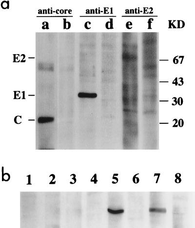
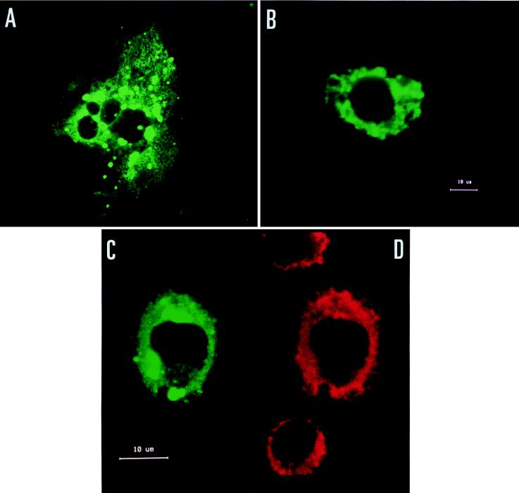
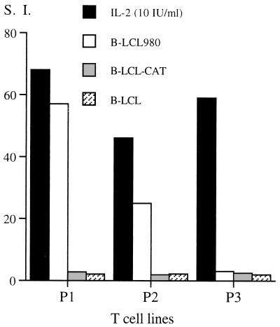
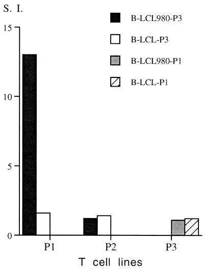
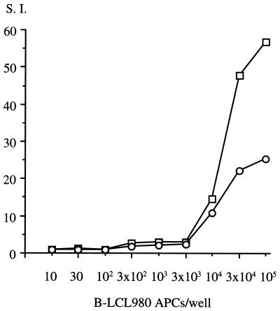
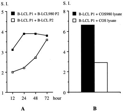
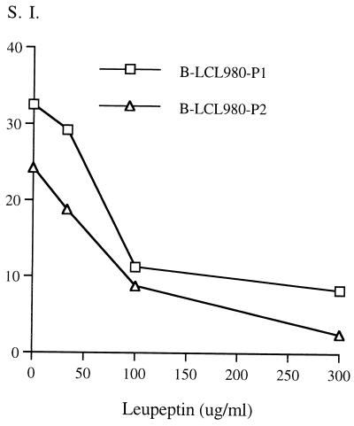
The induction of an efficient CD4(+) T-cell response against hepatitis C virus (HCV) is critical for control of the chronicity of HCV infection. The ability of HCV structural protein endogenously expressed in an antigen-presenting cell (APC) to be presented by class II major histocompatibility complex molecules to CD4(+) T cells was investigated by in vitro culture analyses using HCV core-specific T-cell lines and autologous Epstein-Barr virus-transformed B-lymphoblastoid cell lines (B-LCLs) expressing structural HCV antigens. The T- and B-cell lines were generated from peripheral blood mononuclear cells derived from HCV-infected patients. Expression and intracellular localization of core protein in transfected cells were determined by immunoblotting and immunofluorescence. By stimulation with autologous B-LCLs expressing viral antigens, strong T-cell proliferative responses were induced in two of three patients, while no substantial stimulatory effects were produced by B-LCLs expressing a control protein (chloramphenicol acetyltransferase) or by B-LCLs alone. The results showed that transfected B cells presented mainly endogenously synthesized core peptides. Presentation of secreted antigens from adjacent antigen-expressing cells was not enough to stimulate a core-specific T-cell response. Only weak T-cell proliferative responses were generated by stimulation with B-LCLs that had been pulsed beforehand with at least a 10-fold-higher amount of transfected COS cells in the form of cell lysate, suggesting that presentation of antigens released from dead cells in the B-LCL cultures had a minimal role. Titrating numbers of APCs, we showed that as few as 10(4) transfected B-LCL APCs were sufficient to stimulate T cells. This presentation pathway was found to be leupeptin sensitive, and it can be blocked by antibody to HLA class II (DR). In addition, expression of a costimulatory signal by B7/BB1 on B cells was essential for T-cell activation.


Figures

FIG. 1
FIG. 1
Immunoblot analysis of the HCV structural proteins. (a) Structural gene products were detected by MAbs against core (lane a), E1 (lane c), and E2 (lane e) proteins, respectively, by using transfected COS cells. The lysate of untransfected COS7 cells was used as the cellular protein control (lanes b, d, and f). Specific bands for E2 (70 kDa), E1 (35 kDa), and core protein (22 kDa) were identified. Molecular size markers are indicated on the right in kilodaltons (KD). (b) Detection of core protein in the supernatant of B-LCL980 P1 culture. Lanes 1 to 4, medium samples over 12, 24, 48, and 72 h of culture (2 × 10⁶ cells/ml); no core-specific band was observed. Lanes 5 and 7, cell lysate of transiently transfected COS cells and B-LCL980-P1, respectively; lanes 6 and 8, negative cell lysate controls.
FIG. 2
FIG. 2
IF analysis of protein expression and localization. Expression of core protein in COS cells (A) and B-LCL980-P1 (B) was probed by a MAb to core protein (H-29). (C and D) Localization of core protein in association with the ER in doubly immunostained B cells as detected by a digoxigenin-labeled human polyclonal antibody to core protein (channel 1, FITC) (C) and a MAb to ER membrane protein (PDI) (channel 2, Texas red) (D).
FIG. 3
FIG. 3
Lymphoproliferative responses of T-cell lines to autologous B-LCL980 APCs. T cells (3 × 10⁴ cells/well) were stimulated for 2 days in the presence of autologous B-LCL APCs (1 × 10⁵ cells/well), of pCMV980- or pcDNA3/CAT-transfected cells, or of untransfected cells, or in the absence of APCs with IL-2 alone. T-cell proliferative responses are expressed as SI. The background level (expressed as mean counts per minute of triplicate cultures ± SD) for each T-cell line was 508 ± 98 (P1), 609 ± 61 (P2), and 569 ± 66 (P3) cpm.
FIG. 4
FIG. 4
T-lymphoproliferative response to allogeneic HLA-compatible B-LCL APCs. B-LCL980-P3 APCs were used to stimulate T-cell lines from patients P1 (HLA compatible) and P2 (HLA incompatible). Similarly, the T-cell line from patient P3 was stimulated by allogeneic B-LCL980 APCs (B-LCL980-P1). Untransfected APCs were included in each experiment as B-LCL controls. T-lymphoproliferative responses were expressed as SI.
FIG. 5
FIG. 5
Efficient presentation of endogenous HCV antigens to autologous T-cell lines. T cells (3 × 10⁴ cells/well) from patients P1 (squares) and P2 (circles) were stimulated with various numbers of autologous B-LCL980 APCs. The response of each T-cell line is expressed as the SI.
FIG. 6
FIG. 6
Bystander presentation played a minimal role. (A) Untransfected B-LCL-P1 APCs were precultured with HLA-incompatible transfected APCs (B-LCL980-P2) for 12, 24, 48, and 72 h. Two hundred thousand cells from the coculture were then used to stimulate autologous T cells (3 × 10⁴ cells/well) for 2 days. (B) Untransfected B-LCL-P1 APCs were prepulsed for 3 days with a cell lysate of pCMV980-transfected or untransfected COS7 cells. One hundred thousand pulsed APCs were then used to stimulate autologous T-cell lines.
FIG. 7
FIG. 7
Inhibition of antigen presentation by leupeptin. B-LCL980 APCs were pretreated with leupeptin at various concentrations as indicated for 24 h prior to the T-cell proliferative assay. One hundred thousand APCs were added to the T-cell culture in the presence of the same concentration of leupeptin that was used for pretreatment.
FIG. 8
FIG. 8
Role of B-LCL surface molecules in endogenous presentation. T-cell lines were stimulated for 2 days with autologous B-LCL980 APCs in the presence of a MAb to HLA class I (W6/32; 1:20), HLA class II (DR) (L243; 1:20), or BB1 (L307.4; 10 μg/ml). Results were expressed as percentages of the control response in the absence of the antibodies. Control responses were 28,819 ± 602 and 15,477 ± 1,130 cpm (means of triplicate cultures ± SDs) for T-cell lines P1 and P2, respectively.

References

    1. Alexander-Miller M A, Leggatt G R, Berzofsky J A. Selective expansion of high- or low-avidity cytotoxic T lymphocytes and efficacy for adoptive immunotherapy. Proc Natl Acad Sci USA. 1996;93:4102–4107. - PMC - PubMed
    1. Alexander-Miller M A, Leggatt G R, Sarin A, Berzofsky J A. Role of antigen, CD8, and CTL avidity in high dose antigen induction of apoptosis of effector CTL. J Exp Med. 1996;184:485–492. - PMC - PubMed
    1. Alter H J, Purcell R H, Shih J W, Melolder J C, Houghton M, Choo Q L. Detection of antibody to hepatitis C virus in prospectively followed transfusion recipients with acute and chronic non-A, non-B hepatitis. N Engl J Med. 1989;321:1494–1500. - PubMed
    1. Arichi, T., et al. Unpublished data.
    1. Bartido S M, Diment S, Reiss C S. Processing of a viral glycoprotein in the endoplasmic reticulum for class II presentation. Eur J Immunol. 1995;25:2211–2219. - PMC - PubMed

Publication types

MeSH terms