. 1998 Oct;72(10):8309-15.
doi: 10.1128/JVI.72.10.8309-8315.1998.

A cluster of latently expressed genes in Kaposi's sarcoma-associated herpesvirus

Affiliations

A cluster of latently expressed genes in Kaposi's sarcoma-associated herpesvirus

D Dittmer et al. J Virol. 1998 Oct.

Abstract

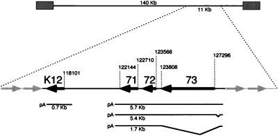
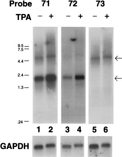
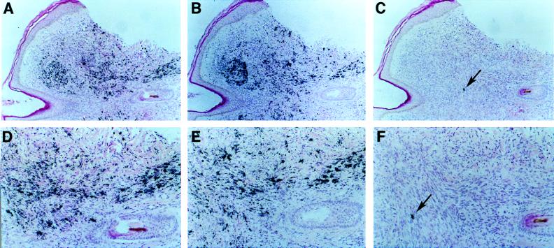
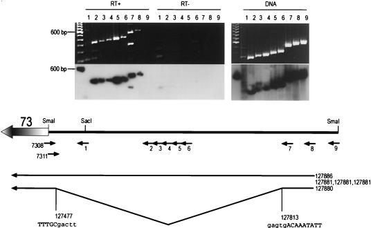
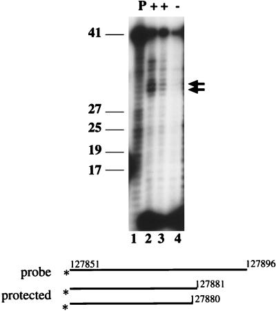
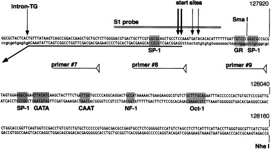
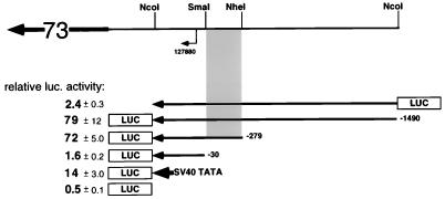
Infection with Kaposi's sarcoma-associated herpesvirus (KSHV) is closely associated with Kaposi's sarcoma (KS) and primary effusion lymphoma, with viral genomes present in a latent state in the majority of tumor cells. Here we describe a cluster of latently expressed viral genes whose mRNAs are generated from a common promoter. Two mRNAs in this region encode the latency-associated nuclear antigen, the product of open reading frame 73 (ORF73). The larger RNA, of 5.8 kb, is an unspliced transcript that includes ORF72 and -71 at its 3' end; it initiates at nucleotides (nt) 127880 to 127886 from a promoter lacking recognizable TATA elements. A less abundant mRNA, of 5.4 kb, is a variant of this transcript, in which 336 nt of 5' noncoding information has been removed by RNA splicing. A third, more abundant RNA is generated from the same promoter region via splicing from the common splice donor at nt 127813 to an acceptor 5' to ORF72; this transcript is the presumed mRNA for ORF72, which encodes the viral cyclin D homolog. All three RNAs are 3' coterminal. In situ hybridization analysis with probes that can detect all three transcripts shows that the RNAs are detectable in a large fraction of BCBL-1 cells prior to lytic induction and in >70% of KS spindle cells in primary KS tumors. This confirms that these transcripts are indeed latent RNAs and suggests a role for their products in viral persistence and/or KSHV-associated proliferation.


Figures

FIG. 1
FIG. 1
Location of clustered latency transcripts in KSHV genome. The numbers indicate nucleotide positions, based on the sequence of Russo et al. (29). The boldface arrows represent ORFK12, -71, -72, and -73.
FIG. 2
FIG. 2
Analysis of LAT transcription. Shown is an autoradiogram of a Northern blot with probes specific for ORF71 (lanes 1 and 2), ORF72 (lanes 2 and 3), and ORF73 (lanes 5 and 6) of BCBL-1 RNA from TPA-induced (lanes 2, 4, and 6) or mock-treated (lanes 1, 3, and 5) cells. The blot was rehybridized with a GAPDH probe as a loading control. The arrows indicate the migration of the 5.8- and 1.7-kb messages.
FIG. 3
FIG. 3
In situ hybridization of a KS tumor section. Shown are subjacent sections of a KS tumor from an HIV-negative patient hybridized with specific probes and counterstained with hematoxylin and eosin stain. Hybridization was done with an ORF72 probe (A and D), an ORFK12 probe (B and E), and a nut-1 probe (C and F). Exposure was for 7 days (A and D) and 3 days (B, E, C, and F). Magnification: ×47 (A to C) and ×96 (D to F). The arrow indicates a single cell positive for the lytic nut-1 RNA.
FIG. 4
FIG. 4
RT-PCR analysis of the ORF73 5′ UTR. BCBL-1 RNA or DNA was reverse transcribed with primer 7308 and PCR amplified with the primers indicated above the lanes. A one hundred-base-pair molecular size ladder is shown in the left lane of each gel. The 600-bp fragment hybridized to the polylinker sequence in the probe. Where indicated (RT−), reverse transcriptase was omitted from the reaction. An ethidium-bromide-stained gel (top) and an autoradiogram of the same gel hybridized with a probe specific for the UTR (lower gel panel) are shown. Note that in the RT+ panel two fragments are amplified in lane 7 and none in lane 9. The deduced structure of the 5′ UTR and the locations of primers (small arrows) are shown below the gels. The numbers on the right of the diagram denote the 5′ end and splice junction, as determined from RACE clones.
FIG. 5
FIG. 5
S1 nuclease protection analysis of the KSHV latency promoter. Shown is an autoradiogram of a 12% denaturing acrylamide gel. Lane 1 shows the input, undigested probe; lanes 2 to 4 show the protected fragments resulting from hybridization to 30 μg of BCBL RNA (lane 2), 15 μg of BCBL-1 RNA (lane 3), or 30 μg of yeast RNA (lane 4) followed by S1 nuclease digestion at 28°C. The arrows point to the two most prominent protected fragments, and size standards are indicated on the left. The asterisks denote the site of the radiolabel.
FIG. 6
FIG. 6
Sequence and putative transcription factors surrounding the latent start site. Shown are the principal start sites (vertical arrows), the predicted binding sites for selected transcription factors (shaded), and the positions of primers used for PCR and S1 analysis. The boldfaced horizontal arrow indicates the 5′ region and splice site of the 5.4- and 1.7-kb transcripts. The boxed sequence represents the predicted ORFK14 coding region. Nucleotide positions are according to the sequence of Russo et al. Lowercase letters represent adjacent sequence not present in the mRNA.
FIG. 7
FIG. 7
Promoter analysis of the KSHV latency promoter. Shown are fold transactivations (averages ± standard deviations) for selected reporter constructs and their locations relative to the latent start site. Nucleotide positions are according to the sequence of Russo et al. (29). The shaded area represents the minimal ORF73 promoter. The boldfaced arrow containing 73 represents the ORF73 transcript. luc., luciferase.

References

    1. Cesarman E, Chang Y, Moore P S, Said J W, Knowles D M. Kaposi’s sarcoma-associated herpesvirus-like DNA sequences in AIDS-related body-cavity-based lymphomas. N Engl J Med. 1995;332:1186–1191. - PubMed
    1. Cesarman E, Nador R G, Bai F, Bohenzky R A, Russo J J, Moore P S, Chang Y, Knowles D M. Kaposi’s sarcoma-associated herpesvirus contains G protein-coupled receptor and cyclin D homologs which are expressed in Kaposi’s sarcoma and malignant lymphoma. J Virol. 1996;70:8218–8223. - PMC - PubMed
    1. Chang Y, Cesarman E, Pessin M S, Lee F, Culpepper J, Knowles D M, Moore P S. Identification of herpesvirus-like DNA sequences in AIDS-associated Kaposi’s sarcoma. Science. 1994;266:1865–1869. - PubMed
    1. Chang Y, Moore P S, Talbot S J, Boshoff C H, Zarkowska T, Godden K, Paterson H, Weiss R A, Mittnacht S. Cyclin encoded by KS herpesvirus. Nature. 1996;382:410. . (Letter.) - PubMed
    1. Church G M, Gilbert W. Genomic sequencing. Proc Natl Acad Sci USA. 1984;81:1991–1995. - PMC - PubMed

Publication types