. 1998 Oct;72(10):8354-7.
doi: 10.1128/JVI.72.10.8354-8357.1998.

The reovirus protein mu2, encoded by the M1 gene, is an RNA-binding protein

Affiliations

The reovirus protein mu2, encoded by the M1 gene, is an RNA-binding protein

L Brentano et al. J Virol. 1998 Oct.

Abstract

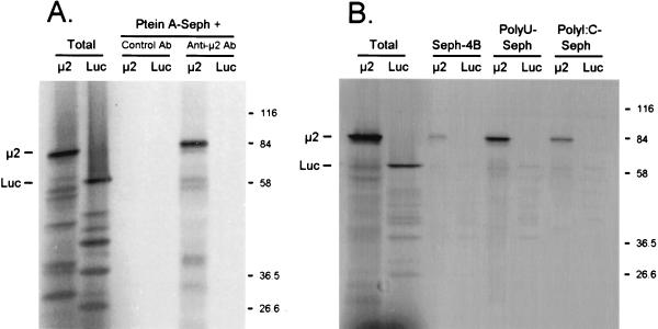
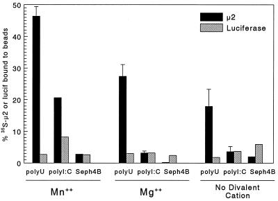
The reovirus M1, L1, and L2 genes encode proteins found at each vertex of the viral core and are likely to form a structural unit involved in RNA synthesis. Genetic analyses have implicated the M1 gene in viral RNA synthesis and core nucleoside triphosphatase activity, but there have been no direct biochemical studies of mu2 function. Here, we expressed mu2 in vitro and assessed its RNA-binding activity. The expressed mu2 binds both poly(I-C)- and poly(U)-Sepharose, and binding activity is greater in Mn2+ than in Mg2+. Heterologous RNA competes for mu2 binding to reovirus RNA transcripts as effectively as homologous reovirus RNA does, providing no evidence for sequence-specific RNA binding by mu2. Protein mu2 is now the sixth reovirus protein demonstrated to have RNA-binding activity.


Figures

FIG. 1
FIG. 1
μ2 binds poly(U)-Sepharose and poly(I-C)–Sepharose. (A) Immunoprecipitations. T7-generated M1 transcripts and control luciferase mRNA (Promega) were translated in rabbit reticulocyte lysates (Promega) containing [35S]Met, and the products were precleared with protein A-Sepharose CL4B (Ptein A-Seph) beads (Pharmacia) that had been washed in TNET buffer (50 mM Tris [pH 8.0], 100 mM NaCl, 5 mM EDTA, 1% Triton X-100). Hyperimmune rabbit antiserum prepared against a serotype 3 Dearing (T3D) μ2-Trp-E fusion protein (32), cross-reactive with T1L-(or 8B)-μ2, was incubated with washed protein A-Sepharose CL4B beads and then resuspended in TNET buffer. Precleared supernatants were then incubated with complexed beads. After extensive washing in radioimmunoprecipitation assay buffer (50 mM Tris [pH 8.0], 100 mM NaCl, 1% Triton X-100, 0.5% deoxycholate, 0.1% SDS), bound protein was eluted by boiling in Laemmli sample buffer (11). Eluted protein and total translated protein were resolved by electrophoresis on a 10% Laemmli SDS-polyacrylamide gel (11), fixed in 5% trichloroacetic acid, dried, and exposed to film. Luc, luciferase; Ab, antibody. (B) RNA binding assays. Sepharose CL4B, poly(I-C)–Sepharose type 6, and poly(U)-Sepharose type 6 (Pharmacia, Piscataway, N.J.) were washed in RNA binding buffer (70 mM NaCl, 10 mM Tris [pH 7.4], 5 mM MnCl2, 1 mM dithiothreitol) and then resuspended in the same buffer. Translated proteins were precleared in Sepharose CL4B, incubated with the indicated beads, and washed in RNA binding buffer. The same gel was used as for panel A.
FIG. 2
FIG. 2
Effects of divalent cations on μ2 binding to poly(U)-Sepharose and poly(I-C)–Sepharose. Sepharose CL4B (Seph4B), poly(U)-Sepharose, and poly(I-C)–Sepharose were washed as for Fig. 1 RNA binding assays, except that 5 mM Mn2+ was substituted for with 5 mM Mg2+ or no divalent cation where indicated. Translated products (as for Fig. 1) were precleared with Sepharose CL4B and then incubated with the indicated beads as for Fig. 1 RNA binding assays, except that all incubations and washes contained the indicated divalent cation. Total translated product and triplicate samples (μ2) or single samples (luciferase [lucif]) bound to the indicated beads were resolved by SDS-PAGE and scanned with a Packard instant imager. The manufacturer’s software was used to select bands of the appropriate molecular weight for quantitation, and the percent of protein bound was calculated relative to total translated μ2 or luciferase (mean ± standard deviation).
FIG. 3
FIG. 3
Protein μ2 is expressed from a recombinant baculovirus. The 8B M1 gene was subcloned into recombinant baculovirus by using the Bac-to-Bac baculovirus expression system (GIBCO BRL, Grand Island, N.Y.). Control GUS-expressing recombinant baculovirus was provided by the manufacturer. T. ni insect cells were infected with virus stock that had been passaged in Sf9 insect cells, and cell cultures were harvested at 48 h (B) or 72 h (A and B) postinfection (POSTINF), washed with phosphate-buffered saline supplemented with 1 mM phenylmethylsulfonyl fluoride, and lysed in radioimmunoprecipitation assay buffer. Lysate supernatants were resolved by electrophoresis on 10% Laemmli SDS–polyacrylamide gels. (A) Coomassie blue staining, duplicate samples. MW, molecular mass (kilodaltons) markers. (B) Western Blot analysis. For Western blot analysis, protein was transferred to Immobilon-P membrane with a semidry blotting system (Millipore, Bedford, Mass.). Detection by the ECL (enhanced chemiluminescence) system (Amersham Life Sciences, Arlington Heights, Ill.) was done according to the manufacturer’s protocol with hyperimmune rabbit antiserum as for Fig. 1 immunoprecipitations.
FIG. 4
FIG. 4
Baculovirus-expressed μ2 binds nucleic acid, and binding is not sequence specific. Lysate supernatants from recombinant baculovirus-infected T. ni cells were incubated with rabbit anti-μ2 antisera and then incubated with protein A-Sepharose CL4B. After extensive washing with radioimmunoprecipitation assay buffer, the Sepharose-protein A-immunocomplexed μ2 or control GUS protein was resuspended in high-salt RNA binding buffer (200 mM NaCl, 30 mM Tris [pH 7.4], 5 mM MnCl2, 0.5 mM dithiothreitol) and incubated with no further addition, with unlabeled (competitor [comp.]) ssDNA (M13mp18; U.S. Biochemical Corp.) or with the indicated quantity of unlabeled (competitor) T7-generated ssRNA transcripts: reovirus positive (pos)- or negative (neg)-strand S4 (18), positive-strand M1, or control feline β-myosin. Triplicate samples were then incubated with T7-generated 32P-labeled ssRNA transcripts as indicated (1 ng per reaction, by extrapolation from the predicted specific activity) and then washed extensively with high-salt RNA binding buffer. Bound RNA was eluted with 1 M NaCl–30 mM Tris (pH 7.4), with 5 mM MnCl2, and electrophoresed on a 1% agarose gel. Gels were acid fixed, dried, and scanned with a Packard instant imager. The manufacturer’s software was used to quantitate bands of the appropriate molecular weight. The percent of 32P-RNA bound was calculated relative to 32P-RNA bound in the absence of competing unlabeled nucleic acid (triplicate samples, mean ± standard error of the mean).

Similar articles

Cited by

References

    1. Boyle J F, Holmes K V. RNA-binding proteins of bovine rotavirus. J Virol. 1986;58:561–568. - PMC - PubMed
    1. Brantley J D, Hunt A G. The N-terminal protein of the polyprotein encoded by potyvirus tobacco vein mottling virus is an RNA-binding protein. J Gen Virol. 1993;74:1157–1162. - PubMed
    1. Burd C G, Dreyfuss G. Conserved structures and diversity of functions of RNA-binding proteins. Science. 1994;265:615–621. - PubMed
    1. Calnan B J, Tidor B, Biancalana S, Hudson D, Frankel A D. Arginine-mediated RNA recognition: the arginine fork. Science. 1991;252:1167–1171. - PubMed
    1. Cleveland D R, Zarbl H, Millward S. Reovirus guanylyltransferase is L2 gene product lambda 2 protein. J Virol. 1986;60:307–311. - PMC - PubMed

Publication types

LinkOut - more resources