. 1998 Oct;72(10):8384-91.
doi: 10.1128/JVI.72.10.8384-8391.1998.

Proviral structure, chromosomal location, and expression of HERV-K-T47D, a novel human endogenous retrovirus derived from T47D particles

Affiliations

Proviral structure, chromosomal location, and expression of HERV-K-T47D, a novel human endogenous retrovirus derived from T47D particles

W Seifarth et al. J Virol. 1998 Oct.

Abstract

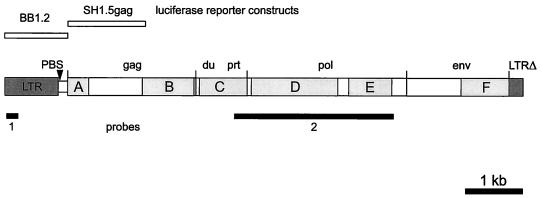
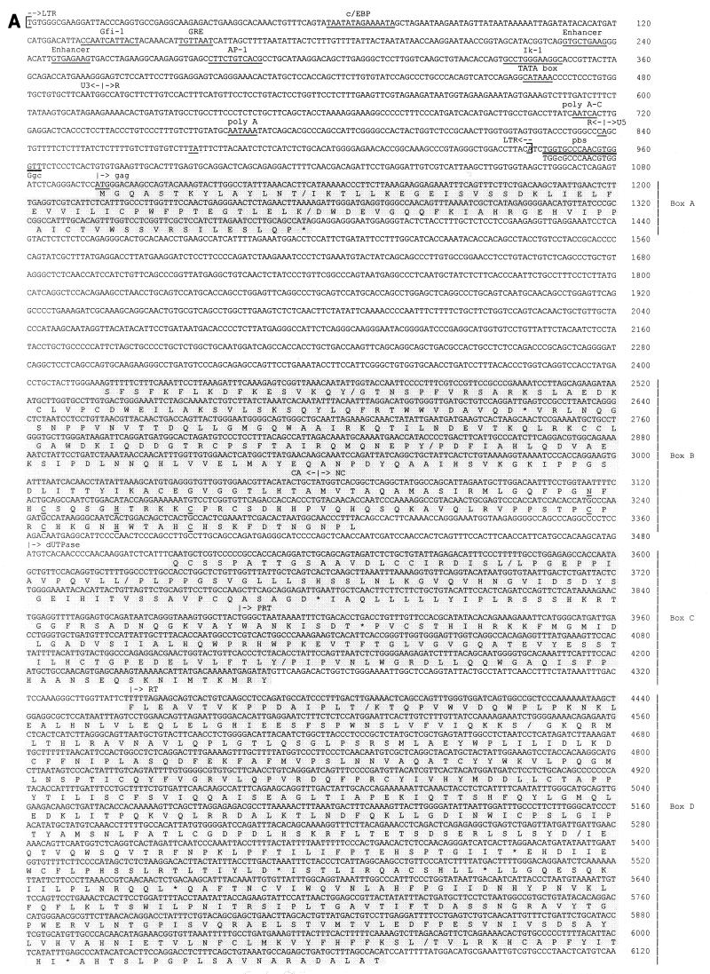
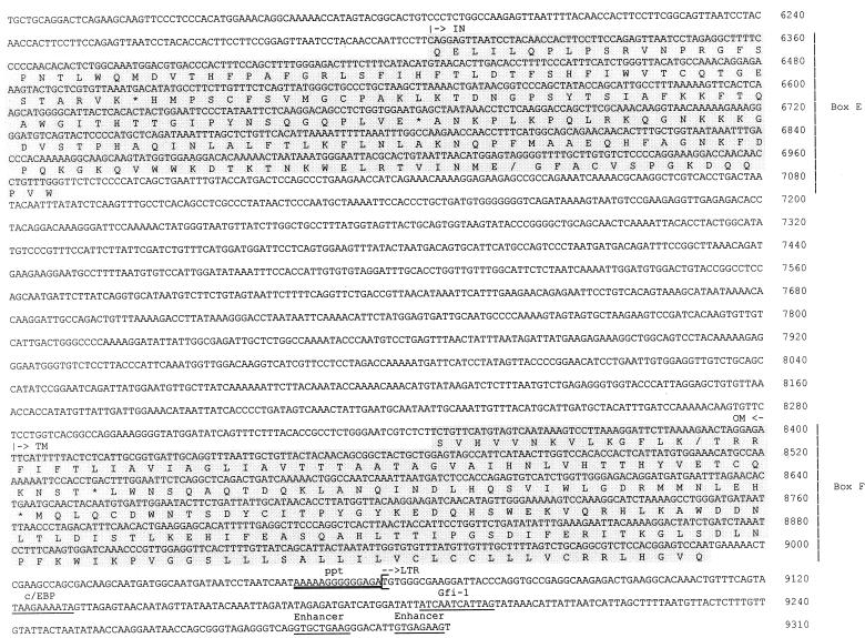
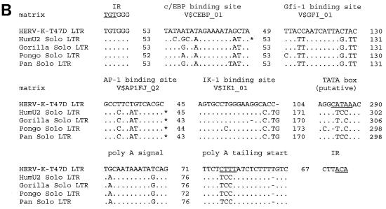
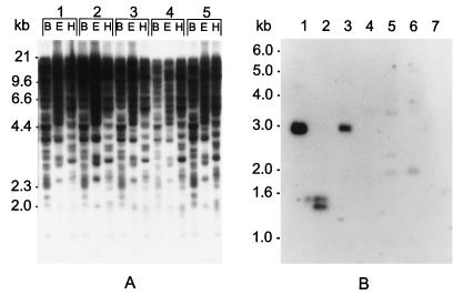
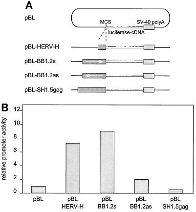
We previously described that type B retrovirus-like particles released from the human mammary carcinoma cell line T47D are pseudotypes and package retroviral RNA of different origins (W. Seifarth, H. Skladny, F. Krieg-Schneider, A. Reichert, R. Hehlmann, and C. Leib-Mösch, J. Virol. 69:6408-6416, 1995). One preferentially packaged retroviral sequence, ERV-MLN, has now been used to isolate the corresponding full-length provirus from a human genomic library. The 9,315-bp proviral genome comprises a complete retroviral structure except for a 3' long terminal repeat (LTR) truncation. A lysine tRNA primer-binding site and phylogenetic analyses assign this human endogenous retroviral element, now called HERV-K-T47D, to the HML-4 subgroup of the HERV-K superfamily. The gag, prt, pol, and env genes exhibit 40 to 60% amino acid identity to HERV-K10. HERV-K-T47D is located on human chromosome 10, with five closely related elements on chromosomes 8, 9, 15, 16, and 19 and several hundred HERV-K-T47D-related solitary LTRs dispersed over the human genome. HERV-K-T47D-related sequences are detected in the genomes of higher primates and Old World monkeys but not in those of New World monkeys. High HERV-K-T47D transcription levels were observed in human placenta tissue, whereas transcription in T47D cells was strictly steroid dependent.


Figures

FIG. 1
FIG. 1
Proviral organization of HERV-K-T47D, locations of hybridization probes, and regions of amino acid identity to HERV-K10. DNA fragments used as hybridization probes are shown as black bars (LTR, probe 1, 229 bp; pol, probe 2, 2.9-kb HindIII-HindIII fragment). Six regions with amino acid identity to HERV-K10 were identified and are depicted as shaded boxes A to F. Abbreviations: du, dUTPase; prt, protease; pol, polymerase; env, envelope. HERV-K-T47D fragments (BB1.2, nt 25 to 1243, 1,218 bp; SH1.5gag, nt 1062 to 2517, 1,455 bp) employed for the construction of recombinant pBL luciferase reporter plasmids used in transient transfection experiments are shown as open bars at the top of the figure. LTRΔ indicates the truncated 3′ LTR.
FIG. 2
FIG. 2
(A) Nucleotide sequence of HERV-K-T47D proviral DNA. LTRs are enclosed in brackets, and the inverted termini TGT and ACA are indicated by arrows. Transcriptional regulatory sequences, i.e., c/EBP, Gfi-1, AP-1, Ik-1, a glucocorticoid-responsive element (GRE), enhancer-like elements, a putative TATA box, a polyadenylation signal, and polyadenylation sites (CA and TA), are underlined once and labeled above. PBS and the polypurine tract (ppt) are double underlined. Sequence complementary to the 3′ end of human lysine tRNA is depicted below the PBS sequence, with lowercase letters being used for mismatches. Translated amino acid sequences with significant homology to HERV-K10 (31), shown under the nucleotide sequence in the six shaded boxes A to F, are those of gag (box A, 40% identity; box B, 49% identity), dUTPase-protease (box C, 59% identity), RT-RNase H (box D, 59% identity), integrase (box E, 59% identity), and env (box F, 58% identity). Frameshifts in the amino acid sequence are indicated with slashes; asterisks correspond to stop codons. Conserved zinc finger motifs (type CX2CX4HX4C) in the NC (box B) region are marked by underlining of the corresponding amino acids. (B) Alignment of putative regulatory elements of the HERV-K-T47D 5′ LTR with corresponding elements from solitary HERV-K-T47D-related LTRs of higher primates (33). Asterisks indicate binding sites which would not have been found with the default parameters of MathInspector (34). However, they were found when a lower threshold was used. Dots and dashes show identical and missing nucleotides, respectively. Under the binding site designations are search string variables used by the program MathInspector. IR, inverted repeat.
FIG. 2
FIG. 2
(A) Nucleotide sequence of HERV-K-T47D proviral DNA. LTRs are enclosed in brackets, and the inverted termini TGT and ACA are indicated by arrows. Transcriptional regulatory sequences, i.e., c/EBP, Gfi-1, AP-1, Ik-1, a glucocorticoid-responsive element (GRE), enhancer-like elements, a putative TATA box, a polyadenylation signal, and polyadenylation sites (CA and TA), are underlined once and labeled above. PBS and the polypurine tract (ppt) are double underlined. Sequence complementary to the 3′ end of human lysine tRNA is depicted below the PBS sequence, with lowercase letters being used for mismatches. Translated amino acid sequences with significant homology to HERV-K10 (31), shown under the nucleotide sequence in the six shaded boxes A to F, are those of gag (box A, 40% identity; box B, 49% identity), dUTPase-protease (box C, 59% identity), RT-RNase H (box D, 59% identity), integrase (box E, 59% identity), and env (box F, 58% identity). Frameshifts in the amino acid sequence are indicated with slashes; asterisks correspond to stop codons. Conserved zinc finger motifs (type CX2CX4HX4C) in the NC (box B) region are marked by underlining of the corresponding amino acids. (B) Alignment of putative regulatory elements of the HERV-K-T47D 5′ LTR with corresponding elements from solitary HERV-K-T47D-related LTRs of higher primates (33). Asterisks indicate binding sites which would not have been found with the default parameters of MathInspector (34). However, they were found when a lower threshold was used. Dots and dashes show identical and missing nucleotides, respectively. Under the binding site designations are search string variables used by the program MathInspector. IR, inverted repeat.
FIG. 3
FIG. 3
(A) Southern blot analysis of human genomic DNA with a probe specific for the HERV-K-T47D LTR (Fig. 1, probe 1). DNA samples (10 μg/lane) from healthy individuals (1 to 5) were restricted to completion, blotted, and hybridized under relaxed stringency conditions (5× SSC, 60°C). Restriction enzymes are abbreviated as follows: B, BamHI; E, EcoRI; and H, HindIII. Marker sizes are indicated on the left. (B) Southern blot analysis of DNA derived from Old World and New World monkeys and higher primates, using a probe specific for the HERV-K-T47D pol gene (Fig. 1, probe 2). High-molecular-weight DNA (10 μg/lane) was restricted to completion with HindIII, blotted, and hybridized under relaxed stringency conditions. The DNAs analyzed were as follows: lane 1, human; lane 2, chimpanzee; lane 3, orangutan; lane 4, Presbytis; lane 5, baboon; lane 6, rhesus monkey; and lane 7, Aotes.
FIG. 4
FIG. 4
HERV-K-T47D transcription in human tissues. (A) Total RNA derived from estradiol- and progesterone-induced (T47D+) or noninduced (T47D−) cells was blotted onto Hybond membranes. All filters were probed with an HERV-K-T47D pol fragment (Fig. 1, probe 2) and washed under conditions of high stringency (0.1× SSC, 65°C). (B) Multiple-tissue Northern blot with 2 μg of mRNA per lane from heart, brain, placenta, lung, liver, skeletal muscle, kidney, and pancreas tissues. To assess RNA quality, the blots (A and B) were rehybridized with a human APC probe and a ubiquitin probe, respectively. Marker sizes are indicated on the left.
FIG. 5
FIG. 5
Analysis of HERV-K-T47D putative LTR promoter activity in T47D cells. (A) pBL-HERV reporter constructs used for luciferase expression assays. The putative LTR 1.2-kb PCR fragment (Fig. 1, fragment BB1.2) was cloned in the sense (pBL-BB1.2s) and the antisense (pBL-BB1.2as) orientations into the luciferase expression vector pBL. As controls, plasmid pBL-SH1.5gag with the insert (SH1.5gag, Fig. 1) and pBL-HERV-H containing the HERV-H LTR promoter of H6 (7) were similarly constructed. MCS, multiple cloning site; SV-40, simian virus 40. (B) Transient expression in T47D cells of HERV-pBL luciferase reporter constructs. T47D cells were transiently transfected according to standard procedures. The luciferase expression driven by the retroviral promoter was measured by a standardized luciferase assay and is shown as bar graphs representing relative promoter activity. All results shown are derived from triplicate experiments.

Similar articles

Cited by

References

    1. Al-Sumidaie A M, Hart C A, Leinster S J, Green C D, McCarthy K. Particles with properties of retroviruses in monocytes from patients with breast cancer. Lancet. 1988;ii:5–9. - PubMed
    1. Boyd M T, Foley B, Brodsky I. Evidence for copurification of HERV-K-related transcripts and a reverse transcriptase activity in human platelets from patients with essential thrombocythemia. Blood. 1997;90:4022–4030. - PubMed
    1. Chen C, Okayama H. High-efficiency transformation of mammalian cells by plasmid DNA. Mol Cell Biol. 1987;7:2745–2752. - PMC - PubMed
    1. Conrad B, Weissmahr R N, Böni J, Arcari R, Schüpbach J, Mach B. A human endogenous retroviral superantigen as candidate autoimmune gene in type I diabetes. Cell. 1997;90:303–313. - PubMed
    1. Cross S H, Charlton J A, Nan X, Bird A P. Purification of CpG islands using a methylated DNA binding column. Nat Genet. 1994;6:236–244. - PubMed

Publication types

Associated data