

Myelin degeneration in multiple system atrophy detected by unique antibodies
- PMID: 9736024
- PMCID: PMC1853025
- DOI: 10.1016/S0002-9440(10)65617-9
Myelin degeneration in multiple system atrophy detected by unique antibodies
Abstract
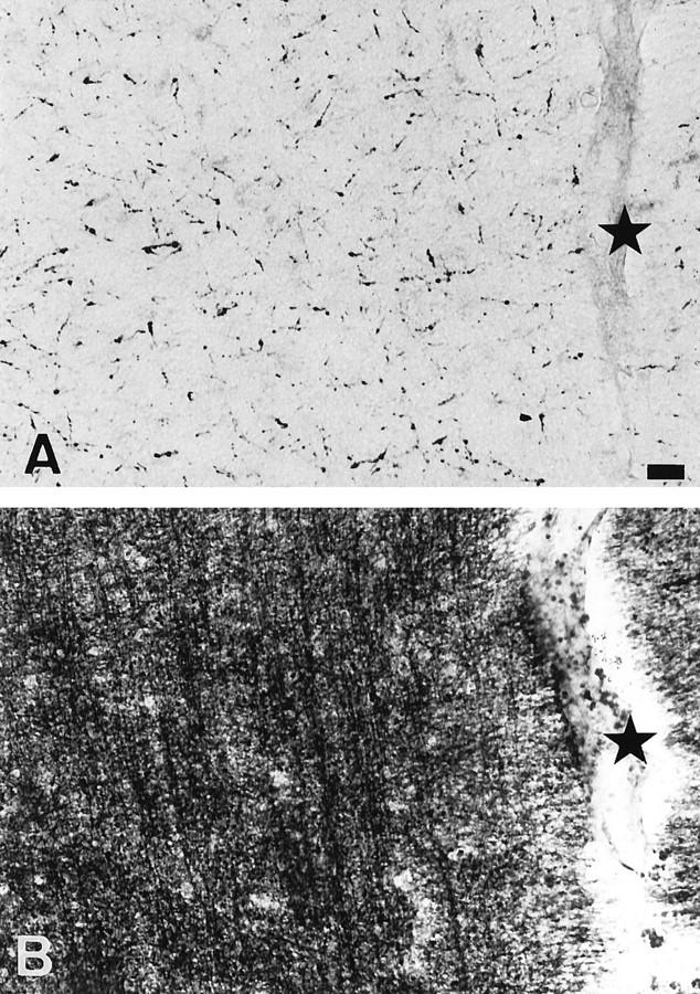
A rabbit antiserum (anti-EP), induced against a synthetic peptide corresponding to residues 68 to 86 of guinea pig myelin basic protein, powerfully immunostained abnormal-appearing oligodendrocytic processes and cell bodies in demyelinating areas associated with multiple system atrophy (MSA). However, as we reported previously, the antiserum, which is highly specific for the sequence QDENPVV corresponding to human myelin basic protein residues 82 to 88, failed to recognize any structures in normal human brain. QD-9, a mouse monoclonal antibody raised against human myelin basic protein residues 69 to 88, which also recognizes specifically the epitope QDENPVV, gave the same results as did anti-EP. The unusual epitope recognized by anti-EP/QD-9 antibodies appears to be accessible in areas of myelin degeneration, and the antibodies have been shown to detect such areas in multiple sclerosis and infarcted brains. These antibodies detect myelin degeneration more widely than previous conventional methods. The present study emphasizes the importance of myelin degeneration in the pathogenesis of multiple system atrophy.
Figures


Comment in
-
Multiple system atrophy: clues from inclusions.Am J Pathol. 1998 Sep;153(3):671-6. doi: 10.1016/S0002-9440(10)65608-8. Am J Pathol. 1998. PMID: 9736015 Free PMC article. Review. No abstract available.
Similar articles
-
Unmasking of an unusual myelin basic protein epitope during the process of myelin degeneration in humans: a potential mechanism for the generation of autoantigens.Am J Pathol. 1997 Apr;150(4):1253-66. Am J Pathol. 1997. PMID: 9094982 Free PMC article.
-
Multiple system atrophy: clues from inclusions.Am J Pathol. 1998 Sep;153(3):671-6. doi: 10.1016/S0002-9440(10)65608-8. Am J Pathol. 1998. PMID: 9736015 Free PMC article. Review. No abstract available.
-
Monoclonal antibodies to distinct regions of human myelin proteolipid protein simultaneously recognize central nervous system myelin and neurons of many vertebrate species.J Neurosci Res. 2006 Feb 15;83(3):415-31. doi: 10.1002/jnr.20748. J Neurosci Res. 2006. PMID: 16416423
-
p25alpha relocalizes in oligodendroglia from myelin to cytoplasmic inclusions in multiple system atrophy.Am J Pathol. 2007 Oct;171(4):1291-303. doi: 10.2353/ajpath.2007.070201. Epub 2007 Sep 6. Am J Pathol. 2007. PMID: 17823288 Free PMC article.
-
[Mechanism of neuronal degeneration of multiple system atrophy].Brain Nerve. 2009 Sep;61(9):1051-60. Brain Nerve. 2009. PMID: 19803404 Review. Japanese.
Cited by
-
Loss of Myelin Basic Protein Function Triggers Myelin Breakdown in Models of Demyelinating Diseases.Cell Rep. 2016 Jul 12;16(2):314-322. doi: 10.1016/j.celrep.2016.06.008. Epub 2016 Jun 23. Cell Rep. 2016. PMID: 27346352 Free PMC article.
-
Multiple system atrophy: a sporadic synucleinopathy.Brain Pathol. 1999 Oct;9(4):721-32. doi: 10.1111/j.1750-3639.1999.tb00553.x. Brain Pathol. 1999. PMID: 10517510 Free PMC article. Review.
-
CSF1R-Mediated Myeloid Cell Depletion Prolongs Lifespan But Aggravates Distinct Motor Symptoms in a Model of Multiple System Atrophy.J Neurosci. 2022 Oct 5;42(40):7673-7688. doi: 10.1523/JNEUROSCI.0417-22.2022. Epub 2022 Sep 6. J Neurosci. 2022. PMID: 36333098 Free PMC article.
-
A brain-targeted, modified neurosin (kallikrein-6) reduces α-synuclein accumulation in a mouse model of multiple system atrophy.Mol Neurodegener. 2015 Sep 23;10:48. doi: 10.1186/s13024-015-0043-6. Mol Neurodegener. 2015. PMID: 26394760 Free PMC article.
-
A historical review of multiple system atrophy with a critical appraisal of cellular and animal models.J Neural Transm (Vienna). 2021 Oct;128(10):1507-1527. doi: 10.1007/s00702-021-02419-8. Epub 2021 Oct 6. J Neural Transm (Vienna). 2021. PMID: 34613484 Free PMC article. Review.
References
-
- Papp MI, Kahn JE, Lantos PL: Glial cytoplasmic inclusions in the CNS of patients with multiple system atrophy (striatonigral degeneration, olivopontocerebellar atrophy and Shy-Drager syndrome). J Neurol Sci 1989, 94:79-100 - PubMed
-
- Chin SSM, Goldman JE: Glial inclusions in CNS degenerative diseases. J Neuropathol Exp Neurol 1966, 55:499-508 - PubMed
-
- Nakazato Y, Yamazaki H, Hirato J, Ishida Y, Yamaguchi H: Oligodendroglial microtubular tangles in olivopontocerebellar atrophy. J Neuropathol Exp Neurol 1990, 49:521-530 - PubMed
-
- Arai N, Nishimura M, Oda M, Morimatsu Y, Ohe R, Nagatomo H: Immunohistochemical expression of microtubule-associated protein 5 (MAP5) in glial cells in multiple system atrophy. J Neurol Sci 1992, 109:102-106 - PubMed
Publication types
MeSH terms
Substances
LinkOut - more resources
Full Text Sources
Molecular Biology Databases
