Comparative Study
. 1998 Sep;153(3):757-65.
doi: 10.1016/S0002-9440(10)65619-2.

Basic fibroblast growth factor is neither necessary nor sufficient for the development of retinal neovascularization

Affiliations
Comparative Study

Basic fibroblast growth factor is neither necessary nor sufficient for the development of retinal neovascularization

H Ozaki et al. Am J Pathol. 1998 Sep.

Abstract

Basic fibroblast growth factor (FGF2) is constitutively expressed in the retina and its expression is increased by a number of insults, but its role in the retina is still uncertain. This study was designed to test the hypothesis that altered expression of FGF2 in the retina affects the development of retinal neovascularization. Mice with targeted disruption of the Fgf2 gene had no detectable expression of FGF2 in the retina by Western blot, but retinal vessels were not different in appearance or total area from wild-type mice. When FGF2-deficient mice were compared with wild-type mice in a murine model of oxygen-induced ischemic retinopathy, they developed the same amount of retinal neovascularization. Transgenic mice with a rhodopsin promoter/Fgf2 gene fusion expressed high levels of FGF2 in retinal photoreceptors but developed no retinal neovascularization or other abnormalities of retinal vessels; in the ischemic retinopathy model, they showed no significant difference in the amount of retinal neovascularization compared with wild-type mice. These data indicate that FGF2 expression is not necessary nor sufficient for the development of retinal neovascularization. This suggests that agents that specifically antagonize FGF2 are not likely to be useful adjuncts in the treatment of retinal neovascularization and therapies designed to increase FGF2 expression are not likely to be complicated by retinal neovascularization.


Figures

Figure 1.
Figure 1.
Schematic map of the rhodopsin/Fgf2 fusion gene. The bovine rhodopsin promoter is the previously described −2174 to +70 fragment. The FGF2 fragment extends from +84 to +1018 of the Fgf2 gene with translation start sites at +280, +322, and +445, and the stop codon at +910 to +912. The oligonucleotide primers used to screen genomic DNA for presence of the transgene are represented by P1 and P2. The primers used for RT-PCR are represented by P3 and P2. The transcription start site is marked +1.
Figure 2.
Figure 2.
Immunoblot of FGF2 in eye extracts from wild-type and FGF2-deficient mice. Lane 1: Human recombinant FGF2 (25 ng). Lane 2: Extract from FGF2-deficient mice. Lane 3: Extract from wild-type mice.
Figure 3.
Figure 3.
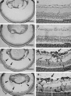
Assessment of retinal blood vessels and neovascularization in wild-type and FGF2-deficient mice with and without ischemic retinopathy. Retinal frozen sections were histochemically stained with the endothelial cell-selective lectin, griffonia simplicifolia I, using the peroxidase-antiperoxidase technique. Retinal blood vessels within the retinas and neovascularization on the surface of the retinas are stained with reaction product, and other retinal cells are counterstained. A and B: Normal retinal vessels in nonischemic P17 wild-type mice. C and D: Nonischemic P17 FGF2-deficient mice also show normal retinal vessels and no identifiable retinal abnormalities. E and F: Wild-type mice with ischemic retinopathy show clumps of endothelial cells on the surface of the retina due to neovascularization. G and H: FGF2-deficient mice with ischemic retinopathy also show numerous endothelial cells on the surface of the retina. Bars: 0.5 mm (A, C, E, and G) and 50 μm (B, D, F, and H).
Figure 4.
Figure 4.
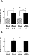
Quantitation of the area of endothelial cell staining in retinal sections of wild-type and FGF2-deficient mice with and without ischemic retinopathy. A: Total area of endothelial cell staining in the retina. Using Student’s unpaired t-test, there is no difference between nonischemic wild-type versus FGF2-deficient mice, nor is there any difference between ischemic wild-type versus FGF2-deficient mice. B: Area of endothelial cell staining on the surface of the retina. Using Student’s unpaired t-test, there is no difference between nonischemic wild-type versus FGF2-deficient mice, nor is there any difference between ischemic wild-type versus FGF2-deficient mice.
Figure 5.
Figure 5.
Assessment of transgene mRNA level in retinal RNA by RT-PCR. Total RNA was extracted from the retina of a transgene-negative mouse (tg(−)) and two transgene-positive mice from five different lines (bF14, bF16, bF19, bF28, and bF8), and RT-PCR was done simultaneously for VEGF transgene mRNA and S16 ribosomal protein mRNA. The size of the band amplified with transgene primers was compatible with the 478 bp predicted for amplification from mRNA. Mice from the same line showed similar expression, and expression was good for all lines except bF16. Mice ranged in age from 2 to 4.5 months.
Figure 6.
Figure 6.
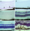
Staining for FGF2 or griffonia simplicifolia I in the retinas of mice overexpressing FGF2 or VEGF. A: A 2-month-old FGF2 transgene-positive mouse shows immunohistochemical staining for FGF2 selectively in retinal photoreceptors. B: Staining is absent on a serial section when nonimmune serum is substituted for primary antibody. C: A transgene-negative mouse shows faint diffuse staining for FGF2 throughout the retina but no selective staining in photoreceptors. D to F: Staining with griffonia simplicifolia I of the retina from a 4-month-old (D) or a 2-month-old (E) FGF2 transgenic mouse shows normal retinal blood vessels and no neovascularization, whereas the retina of a 1-month-old VEGF transgenic mouse shows a large clump of neovascularization F, (arrows). Bar: 50 μm.
Figure 7.
Figure 7.
Quantitation of the area of endothelial cell staining in retinal sections of wild-type or FGF2 transgenic mice with and without ischemic retinopathy. N = 7 for each group. Using Student’s unpaired t-test, there is no significant difference between ischemic wild-type versus ischemic FGF2 transgenic mice.

Comment in

Similar articles

Cited by

References

    1. Kahn H, Hiller R: Blindness caused by diabetic retinopathy. Am J Ophthalmol 1974, 78:58-67 - PubMed
    1. Michaelson I: The mode of development of the vascular system of the retina with some observations on its significance for certain retinal diseases. Trans Ophthalmol Soc UK 1948, 68:137-180
    1. Ashton N: Retinal vascularization in health and disease. Am J Ophthalmol 1957, 44:7-17 - PubMed
    1. Shweiki D, Itin A, Soffer D, Keshet E: Vascular endothelial growth factor induced by hypoxia may mediate hypoxia-initiated angiogenesis. Nature 1992, 359:843-845 - PubMed
    1. Plate KH, Breier G, Welch HA, Risau W: Vascular endothelial growth factor is a potential tumor angiogenesis factor in human gliomas in vivo. Nature 1992, 359:845-848 - PubMed

Publication types

MeSH terms

LinkOut - more resources