Expression of human herpesvirus-6 antigens in benign and malignant lymphoproliferative diseases
- PMID: 9736030
- PMCID: PMC1853007
- DOI: 10.1016/S0002-9440(10)65623-4
Expression of human herpesvirus-6 antigens in benign and malignant lymphoproliferative diseases
Abstract
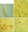
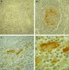
Immunohistochemistry was used to look for the expression of human herpesvirus-6 (HHV-6) antigens in a well characterized series of benign, atypical, and malignant lymphoid lesions, which tested positive for the presence of HHV-6 DNA. A panel of specific antibodies against HHV-6 antigens, characteristic either of the early (p41) or late (p101K, gp106, and gp116) phases of the viral cycle, was applied to the lymphoid tissues from 15 non-Hodgkin's lymphomas, 14 Hodgkin's disease cases, 5 angioimmunoblastic lymphadenopathies with dysproteinemia, 14 reactive lymphadenopathies, and 2 cases of sinus histiocytosis with massive lymphadenopathy (Rosai-Dorfman disease). In lymphomatous tissues, the expression of late antigens was documented only in reactive cells, and mainly in plasma cells. Of interest, the expression of the early p41 antigen was detected in the so-called "mummified" Reed-Sternberg cells, in two Hodgkin's disease cases. In reactive lymphadenopathies, the HHV-6 late antigen-expressing cells were plasma cells, histiocytes, and rare granulocytes distributed in interfollicular areas. In both cases of Rosai-Dorfman disease, the p101K showed an intense staining in follicular dendritic cells of germinal centers, whereas the gp106 exhibited an intense cytoplasmic reaction in the abnormal histiocytes, which represent the histological hallmark of the disease. The expression of HHV-6 antigens is tightly controlled in lymphoid tissues. The lack of HHV-6 antigen expression in neoplastic cells and the limited expression in degenerating Reed-Sternberg cells argue against a major pathogenetic role of the virus in human lymphomagenesis. The detection of a rather unique pattern of viral late antigen expression in Rosai-Dorfman disease suggests a possible pathogenetic involvement of HHV-6 in some cases of this rare lymphoproliferative disorder.
Figures


Similar articles
-
Semi-quantitative in situ hybridization and immunohistology for antigen expression of human herpesvirus-6 in various lymphoproliferative diseases.In Vivo. 1994 Jul-Aug;8(4):517-26. In Vivo. 1994. PMID: 7893978
-
Human herpesvirus-6 (HHV-6) in Hodgkin's disease: cellular expression of viral antigens as compared to oncogenes met and fes, tumor suppressor gene product p53, and interleukins 2 and 6.In Vivo. 1994 Jul-Aug;8(4):501-16. In Vivo. 1994. PMID: 7893977
-
Human herpesvirus 6 in human immunodeficiency virus-infected individuals: association with early histologic phases of lymphadenopathy syndrome but not with malignant lymphoproliferative disorders.J Med Virol. 1996 Apr;48(4):344-53. doi: 10.1002/(SICI)1096-9071(199604)48:4<344::AID-JMV8>3.0.CO;2-7. J Med Virol. 1996. PMID: 8699167
-
The new lymphotropic herpesviruses (HHV-6, HHV-7, HHV-8) and hepatitis C virus (HCV) in human lymphoproliferative diseases: an overview.Haematologica. 1996 May-Jun;81(3):265-81. Haematologica. 1996. PMID: 8767534 Review.
-
Histological diversity of reactive and atypical proliferative lymph node lesions in systemic lupus erythematosus patients.Pathol Res Pract. 2007;203(6):423-31. doi: 10.1016/j.prp.2007.03.002. Epub 2007 May 30. Pathol Res Pract. 2007. PMID: 17540509 Review.
Cited by
-
Facial cutaneous Rosai-Dorfman disease: A case report and literature review.Exp Ther Med. 2015 Apr;9(4):1389-1392. doi: 10.3892/etm.2015.2260. Epub 2015 Feb 5. Exp Ther Med. 2015. PMID: 25780440 Free PMC article.
-
Rosai-Dorfman disease with renal involvement and associated autoimmune haemolytic anaemia in a 12-year-old girl: A case report.BMC Pediatr. 2020 Oct 8;20(1):470. doi: 10.1186/s12887-020-02368-3. BMC Pediatr. 2020. PMID: 33032570 Free PMC article.
-
Rosai-dorfman disease in thoracic spine: a rare case of compression fracture.Korean J Spine. 2014 Sep;11(3):198-201. doi: 10.14245/kjs.2014.11.3.198. Epub 2014 Sep 30. Korean J Spine. 2014. PMID: 25346769 Free PMC article.
-
Rosai-Dorfman disease in 6-year-old child: Presentation, diagnosis, and treatment.Clin Case Rep. 2021 Sep 21;9(9):e04795. doi: 10.1002/ccr3.4795. eCollection 2021 Sep. Clin Case Rep. 2021. PMID: 34584701 Free PMC article.
-
Mutually exclusive recurrent KRAS and MAP2K1 mutations in Rosai-Dorfman disease.Mod Pathol. 2017 Oct;30(10):1367-1377. doi: 10.1038/modpathol.2017.55. Epub 2017 Jun 30. Mod Pathol. 2017. PMID: 28664935 Free PMC article.
References
-
- Yamanishi K, Okuno T, Shiraki T, Takahashi M, Kondo T, Asano Y, Kurata T: Identification of human herpesvirus-6 as a causal agent for exanthem subitum. Lancet 1988, i:1065-1067 - PubMed
-
- Pruksananonda P, Breese-Hall C, Insel RA, McIntyre K, Pellett PE, Long CE, Schnabel KC, Pincus PH, Stamey FR, Dambaugh TR, Stewart JA: Primary human herpesvirus 6 infection in young children. N Engl J Med 1992, 326:1445-1450 - PubMed
-
- Merelli E, Sola P, Barozzi P, Torelli G: An encephalitic episode in a multiple sclerosis patient with human herpesvirus 6 latent infection. J Neurol Sci 1996, 137:42-46 - PubMed
Publication types
MeSH terms
Substances
LinkOut - more resources
Full Text Sources

