. 1998 Sep;153(3):899-909.
doi: 10.1016/S0002-9440(10)65631-3.

Radiation-induced p53 and p21WAF-1/CIP1 expression in the murine intestinal epithelium: apoptosis and cell cycle arrest

Affiliations

Radiation-induced p53 and p21WAF-1/CIP1 expression in the murine intestinal epithelium: apoptosis and cell cycle arrest

J W Wilson et al. Am J Pathol. 1998 Sep.

Abstract

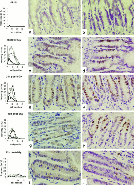
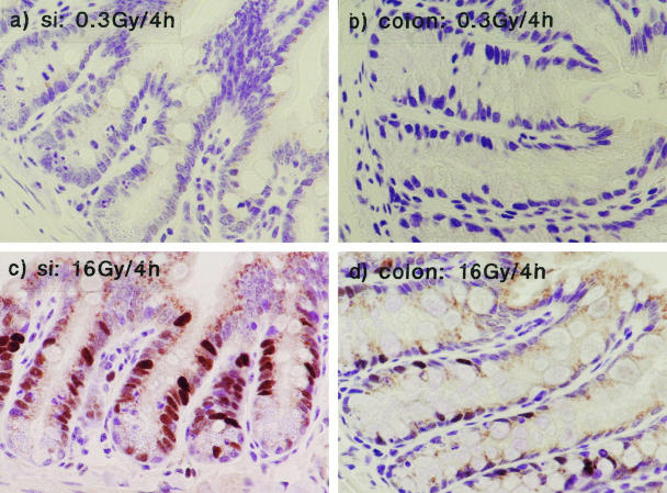
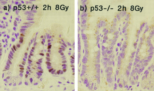
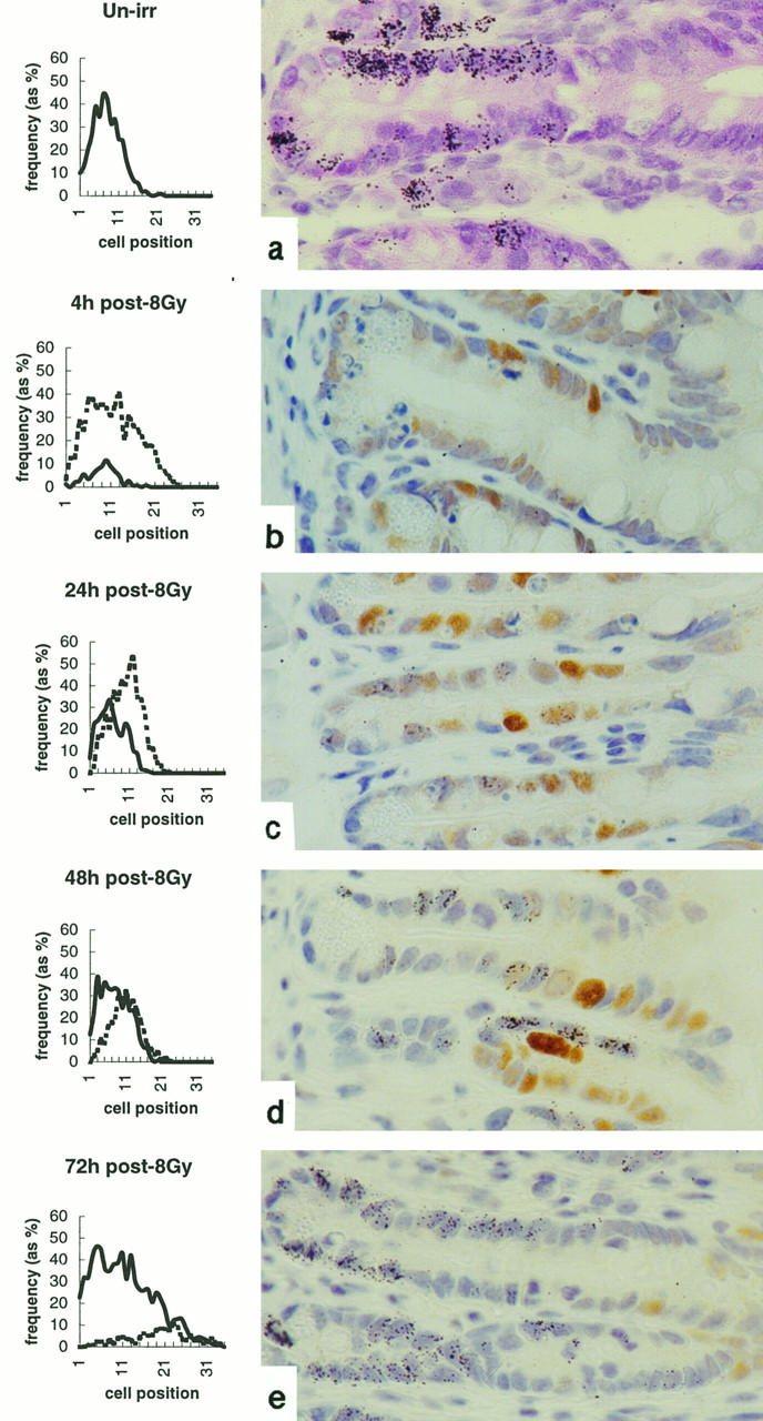
p53-dependent expression of p21WAF-1/CIP1 has been studied in murine intestinal epithelium after exposure to ionizing radiation. In un-irradiated small intestine, neither p53 nor p21WAF-1/CIP1 could be detected by immunohistochemistry. After irradiation (8 Gy), there was a time- and dose-dependent increase in the expression of both proteins. In the small bowel, the positional expression of p53 and p21WAF-1/CIP1 was similar but not coincident. Both proteins could be observed throughout the crypts with greatest frequency of expression over the first 15 cell positions, which includes the stem cell population (approximately positions 3 to 5) and the proliferating, transit cell population (approximately positions 5 to 15). p53-positive cells were primarily distributed toward the base of the crypt relative to p21WAF-1/CIP1. Subdivision of the p53-positive cell population revealed that the cells with strongest p53 immunoreactivity were positioned farther toward the base of the crypt, and their distribution was approximately coincident with the frequency distribution of apoptotic cells. Cells that were either weakly or moderately immunoreactive for p53 were located toward the middle of the crypt and were approximately coincident with the distribution of p21WAF-1/CIP1. The numbers of both p53- and p21WAF-1/CIP1-positive cells declined steadily with time, and by 6 days after irradiation there were very few immunoreactive cells to observe. Radiation-induced increase in p53 and p21WAF-1/CIP1 expression was not detected in mice homozygously null for p53. Expression of p21WAF-1/CIP1 and incorporation of tritiated thymidine were found to be mutually exclusive. In the large bowel, p21WAF-1/CIP1 and p53 expression were observed along the entire length of the colonic crypts after irradiation (8 Gy), and, unlike in the small intestine, this expression was not only maintained but increased over 72 hours. p21WAF-1/CIP1 immunoreactivity was detected in large intestine epithelium up to 6 days after irradiation. The differential expression of p21WAF-1/CIP1, observed between the large and small bowel and within the small intestinal crypts, is discussed.


Figures

Figure 1.
Figure 1.
Radiation-induced apoptosis and expression of p53 and p21WAF-1/CIP1 in murine small intestinal epithelium. Line graphs show distributions of apoptotic (bold, solid line), p53-positive (shaded line), and p21WAF-1/CIP1-positive (dashed line) cells in small intestinal crypts at indicated times after exposure to 8 Gy γ-radiation. Cells are scored on a positional basis, as previously described (Ijiri and Potten 1983). a, c, e, g, and i illustrate p53 immunoreactivity; b, d, f, h, and j show P21WAF-1/CIP1 immunoreactivity. Data are mean results from a minimum of three mice at each time point. At least 1000 cells (50 half-crypts) were scored from each mouse. The data are from one representative experiment typical of three.
Figure 2.
Figure 2.
Relationship between weak and strong p53 and p21WAF-1/CIP1 expression and apoptosis. Shown is the subdivision of the p53-positive cell population into weakly/moderately immunoreactive (shaded line) and strongly immunoreactive (solid, bold line) groups. Also shown are the distribution of apoptotic cells (solid, fine line) and p21WAF-1/CIP1-positive cells (dashed line). Data are for the 4-hour time point after exposure of BDF-1 mice to 8 Gy γ-radiation. Data are mean results from a minimum of three mice at each time point. At least 1000 cells (50 half-crypts) were scored from each mouse. The data are from one representative experiment typical of three.
Figure 3.
Figure 3.
The effect of radiation dose on p21WAF-1/CIP1 expression. a and b: p21WAF-1/CIP1 expression in small and large bowel, respectively, after 0.3 Gy. c and d: p21WAF-1/CIP1 expression after 16 Gy.
Figure 4.
Figure 4.
Radiation-induced p21WAF-1/CIP1 expression is dependent on p53 function. This figure shows p21WAF-1/CIP1 immunoreactivity in wild-type mice (a) or mice homozygously null for p53 (b) 2 hours after exposure to 8 Gy γ-radiation.
Figure 5.
Figure 5.
Western blot demonstrating radiation-induced increase in p21WAF-1/CIP1 expression in the small intestine of mice that are either wt or homozygously null for p53. Figures along the top indicate time (hours) after irradiation (8 Gy). Western samples were prepared by pooling epithelial cell preparations from at least three mice.
Figure 6.
Figure 6.
Radiation-induced apoptosis and expression of p53 and p21WAF-1/CIP1 in murine large intestinal epithelium. Line graphs show distributions of apoptotic cells (bold, solid line), p53-positive cells (shaded line), and p21WAF-1/CIP1 in large intestinal crypts, at indicated times, after exposure to 8 Gy γ-radiation. Cells are scored on a positional basis, as previously described (Ijiri and Potten 1983). a, c, e, g, and i illustrate p53-immunoreactivity; b, d, f, h, and j show p21WAF-1/CIP1 immunoreactivity. Data are mean results from a minimum of three mice at each time point. At least 1000 cells (50 half-crypts) were scored from each mouse. The data are from one representative experiment typical of three.
Figure 7.
Figure 7.
Comparison of the longevity of radiation-induced p21WAF-1/CIP1 expression in the small and large bowel. Shown are large bowel (a and c) and small bowel (b and d) at either 96 hours (a and b) or 6 days (c and d) after irradiation. Data are the mean results from a minimum of four mice at each time point. At least 1000 cells (50 half-crypts) were scored from each mouse. The data are from one experiment typical of two.
Figure 8.
Figure 8.
Effect of radiation on [3H]thymidine incorporation and its relationship to p21WAF-1/CIP1 expression in the small bowel. Line graphs show the distribution of thymidine-labeled (solid line) and p21WAF-1/CIP1-positive (dashed line) cells at indicated times after exposure to 8 Gy γ-radiation. Accompanying plates demonstrate more clearly the distribution of thymidine incorporation (as black silver grains) and p21WAF-1/CIP1 immunoreactivity. Data are mean results from a minimum of three mice at each time point. At least 1000 cells (50 half-crypts) were scored from each mouse. The data are from one experiment typical of two.
Figure 9.
Figure 9.
Effect of radiation on [3H]thymidine incorporation and its relationship to p21WAF-1/CIP1 expression in the large bowel. Line graphs show the distribution of thymidine-labeled (solid line) and p21WAF-1/CIP1-positive (dashed line) cells at indicated times after exposure to 8 Gy γ-radiation. Accompanying plates demonstrate more clearly the distribution of thymidine incorporation (as black silver grains) and p21WAF-1/CIP1 immunoreactivity. Data are mean results from a minimum of three mice at each time point. At least 1000 cells (50 half-crypts) were scored from each mouse. The data are from one experiment typical of two.

Similar articles

Cited by

References

    1. Jackson SP: The recognition of DNA damage. Curr Opin Genet Dev 1996, 6:19-25 - PubMed
    1. Kastan MB, Onyckwere O, Sidransky D, Vogelstein B, Craig RW: Participation of p53 protein in the cellular response to DNA damage. Cancer Res 1991, 51:6304-6311 - PubMed
    1. Kastan MB, Zhan Q, El-Deiry WS, Carrier F, Jacks T, Walsh WV, Plunkett BS, Vogelstein B, Fornace AJ, Jr: A mammalian cell cycle checkpoint pathway utilizing p53 and GADD45 is defective in ataxia telangiectasia. Cell 1992, 71:587-597 - PubMed
    1. Wood RD: DNA repair in eukaryotes. Annu Rev Biochem 1996, 65:135-167 - PubMed
    1. Reed JC: Double identity for proteins of the Bcl-2 family. Nature 1997, 387:773-776 - PubMed

Publication types

MeSH terms