Comparative Study
. 1998 Sep;153(3):945-54.
doi: 10.1016/S0002-9440(10)65636-2.

Differential expression and origin of membrane-type 1 and 2 matrix metalloproteinases (MT-MMPs) in association with MMP2 activation in injured human livers

Affiliations
Comparative Study

Differential expression and origin of membrane-type 1 and 2 matrix metalloproteinases (MT-MMPs) in association with MMP2 activation in injured human livers

N Théret et al. Am J Pathol. 1998 Sep.

Abstract

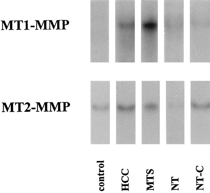
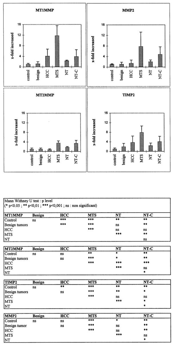
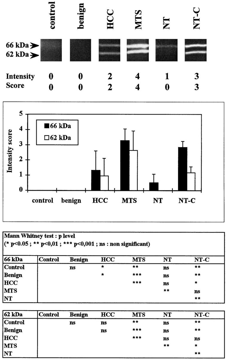
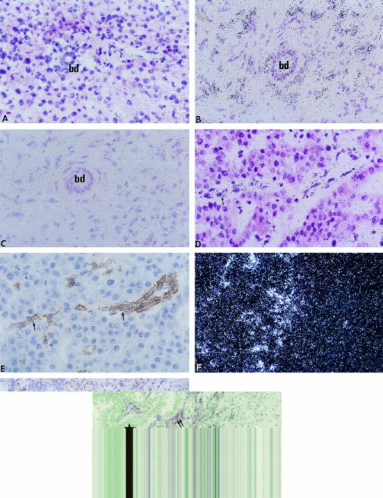
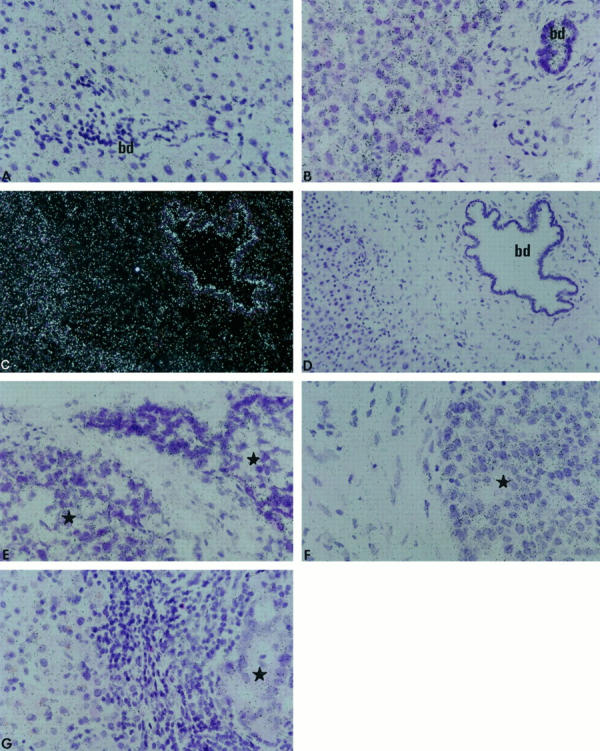
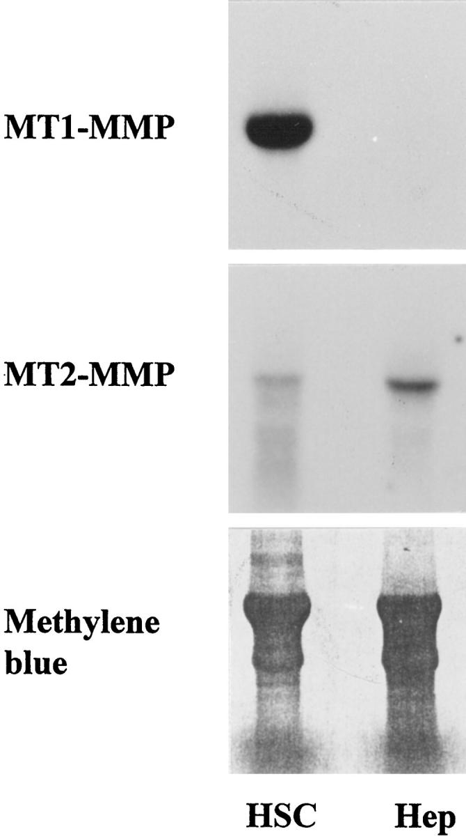
Matrix metalloproteinase-2 (MMP2) activation is associated with basement membrane remodeling that occurs in injured tissues and during tumor invasion. The newly described membrane-type MMPs (MT-MMPs) form a family of potential MMP2 activators. We investigated the localization and steady-state levels of MT1-MMP and MT2-MMP mRNA, compared with those of MMP2 and tissue inhibitor of MMP-2 in 22 hepatocellular carcinomas, 12 liver metastases from colonic adenocarcinomas, 13 nontumoral samples from livers with metastases, 10 benign tumors, and 6 normal livers. MMP2 activation was analyzed by zymography in the same series. The expression of MT1-MMP mRNA and the activation of MMP-2 were increased in hepatocellular carcinomas, metastases, and cholestatic nontumoral samples. MT2-MMP mRNA was rather stable in the different groups. MT1-MMP mRNA levels, but not MT2-MMP mRNA, correlated with MMP-2 and tissue inhibitor of MMP-2 mRNA levels and with MMP2 activation. In situ hybridization showed that MT1-MMP mRNA was expressed in stromal cells, and MT2-MMP mRNA was principally located in both hepatocytes and biliary epithelial cells. Consistently, freshly isolated hepatocytes expressed only MT2-MMP mRNA, and culture-activated hepatic stellate cells showed high levels of MT1-MMP mRNA. These results indicate that in injured livers, MMP2 activation is related to a coordinated high expression of MMP2, tissue inhibitor of MMP-2, and MT1-MMP. Furthermore, the finding of a preferential expression of MT2-MMP in hepatocytes, together with our previous demonstration that the activation of stellate cell-derived MMP2 in co-culture requires interactions with hepatocytes (Am J Pathol 1997, 150:51-58), suggests that parenchymal cells might play a pivotal role in the MMP2 activation process.


Figures

Figure 1.
Figure 1.
Northern blot analysis of MT1-MMP and MT2-MMP mRNA in human livers. Total RNAs were extracted from liver biopsies, resolved by electrophoresis onto agarose gels, and blotted onto Hybond N nylon. MTS, hepatic metastasis from colonic adenocarcinomas; NT, nontumorous samples from livers with metastases (with either minimal histopathological changes (NT) or cholestasis (NT-C).
Figure 2.
Figure 2.
Dot blot analysis of the steady-state MT1-MMP, MT2-MMP, MMP2, and TIMP2 mRNA levels in control livers, benign tumors, HCCs, hepatic metastases from colonic adenocarcinomas (MTS), and nontumorous samples from livers with metastases with either minimal histopathological changes (NT) or cholestasis (NT-C). Top: Densitometric values from each probe within the linear range were normalized with the 18S oligoprobe and plotted. Columns, mean; bars, SD. Bottom: The Mann-Whitney U test was used to test the significance of the differences between means.
Figure 3.
Figure 3.
Zymography analyses. Sections (5 μm/1 cm2) from 53 biopsy samples (control livers, benign tumors, HCCs, hepatic metastases from colonic adenocarcinomas (MTS), and nontumorous samples from livers with metastases with either minimal histopathological changes (NT) or cholestasis (NT-C) were homogenized in sample buffer and electrophoresed on an SDS-7% polyacrylamide gel copolymerized with 1 mg/ml gelatin. Five intensity levels based on densitometric analyses were determined for the Mr 66 and Mr 62 kd bands. Top: Representative zymogram with densitometric range. Middle: Semiquantitative analyses of gelatinolytic activity. Bottom: Results are expressed as means ± SD. The Mann-Whitney U test was used to test the significance of the differences between means.
Figure 4.
Figure 4.
In situ hybridization of MT1-MMP mRNA in normal liver (A), cholestatic liver (B), HCC (D), and metastasis from a colonic adenocarcinoma (F). Controls include an immunohistochemical detection of von Willebrand factor in HCC (E, section contiguous to D) and α-smooth muscle actin in metastasis (G, section contiguous to F) and in situ hybridization with the sense probe (C, section contiguous to B). bd, bile duct; star, tumor; arrows, endothelial cells; double arrows, myofibroblasts. Bright-field (A to D) and dark-field (F) photomicrographs of autoradiographs stained with hematoxylin and eosin. Magnification: A to E, ×400; F and G, ×200.
Figure 5.
Figure 5.
In situ hybridization of MT2-MMP mRNA in normal liver (A), cholestatic liver (B and C), HCCs (E and F), and metastasis from a colonic adenocarcinoma (G). Control with sense probe in situ hybridization is shown (D, section contiguous to C). bd, bile duct; star, tumor. Bright-field (A, B, and D to G) and dark-field (C) photomicrographs of autoradiographs stained with hematoxylin and eosin. Magnification: A, B, and E to G, ×400; C and D, ×200.
Figure 6.
Figure 6.
Northern blots of MT1-MMP and MT2-MMP mRNA in culture-activated stellate cells at passage 6 (HSC) and in freshly isolated hepatocytes (Hep). Note the strong expression of MT1-MMP mRNA in stellate cells, which contrasts with the low levels of MT2-MMP mRNA. Hepatocytes are positive for MT2-MMP mRNA only. Bottom: Transferred RNA stained with methylene blue.

References

    1. Matrisian LM: Metalloproteinases and their inhibitors in matrix remodelling. Trends Genet 1990, 6:121-125 - PubMed
    1. Stetler-Stevenson WG, Aznavoorian S, Liotta LA: Tumor cell interactions with the extracellular matrix during invasion and metastasis. Annu Rev Cell Biol 1993, 9:541-573 - PubMed
    1. Birkedal-Hansen H: Proteolytic remodeling of extracellular matrix. Curr Opin Cell Biol 1995, 7:728-735 - PubMed
    1. Corcoran ML, Hewitt RE, Kleiner DE, Jr, Stetler-Stevenson WG: MMP-2: expression, activation and inhibition. Enzyme Protein 1996, 49:7-19 - PubMed
    1. Yu AE, Hewitt RE, Kleiner DE, Stetler-Stevenson WG: Molecular regulation of cellular invasion: role of gelatinase A and TIMP-2. Biochem Cell Biol 1996, 74:823-831 - PubMed

Publication types

MeSH terms

Substances