

Microdissecting the genetic events in nephrogenic rests and Wilms' tumor development
- PMID: 9736048
- PMCID: PMC1853014
- DOI: 10.1016/S0002-9440(10)65641-6
Microdissecting the genetic events in nephrogenic rests and Wilms' tumor development
Abstract
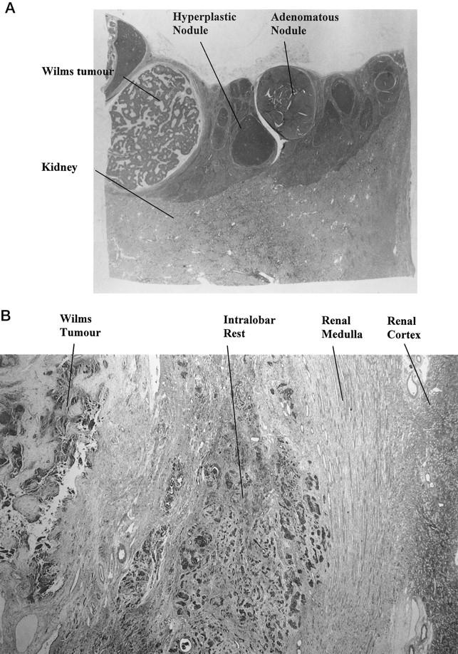
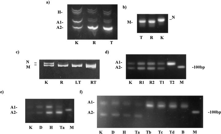
Nephrogenic rests are precursor lesions associated with about 40% of Wilms' tumors. This study identifies genetic steps occurring in the development of Wilms' tumor. Thirty-four Wilms' tumors with nephrogenic rests and/or areas of anaplasia were microdissected from paraffin sections to determine whether and at what stage loss of heterozygosity (LOH) occurred, using polymerase chain reaction-based polymorphic markers at 11p13, 11p15, and 16q. LOH at these loci have been identified in Wilms' tumors and are associated with identified or putative tumor suppressor genes. Three cystic nephromas/cystic partially differentiated nephroblastomas were also examined. LOH was detected in six cases at 11p13 and in six cases at 11p15, and two of these cases had LOH at both loci. All intralobar rests showing LOH also showed LOH in the tumor. A case with a small perilobar rest showed LOH of 11p13 only in the tumor. Five cases showing LOH at 16q were identified (this was identified only in the tumor, and not in the associated rest), and three of these had recurrence of the tumor. Two cases had a WT1 mutation (one germline and the other somatic), as well as LOH in both the intralobar rest and the tumor. A cystic partially differentiated nephroblastoma showed loss at 11p13 and 11p15, as well as at 16q. This study suggests that LOH at 11p13 and 11p15 and WT1 mutations are early events but that LOH at 16q occurs late in the pathogenesis of Wilms' tumor. Intralobar and perilobar nephrogenic rests are known to have different biological behaviors, and this study suggests that they are genetically different. A multistep model of Wilms' tumor pathogenesis is supported by these findings.
Figures

References
-
- Murphy WM, Beckwith JB, Farrow GM: Tumors of the kidney. Atlas of Tumor Pathology: Tumors of the Kidney, Bladder, and Related Urinary Structures. 1994:pp 1-192 Armed Forces Institute of Pathology, Washington DC
-
- Bove KE, McAdams AJ: Multifocal nephroblastic neoplasia. J Natl Cancer Inst 1978, 61:285-294 - PubMed
-
- Beckwith JB, Kiviat NB, Bonadio JF: Nephrogenic rests, nephroblastomatosis, and the pathogenesis of Wilms’ tumour. Pediatr Pathol 1990, 10:1-36 - PubMed
-
- Bard JBL, McConnell JE, Davies JA: Towards a genetic basis for kidney development. Mech Dev 1994, 48:3-11 - PubMed
-
- Chao LY, Huff V, Tomlinson G, Riccardi VM, Strong LC, Saunders GF: Genetic mosaicism in normal tissues of Wilms’ tumour patients. Nat Genet 1993, 3:127-131 - PubMed
Publication types
MeSH terms
Substances
LinkOut - more resources
Full Text Sources
Medical
