. 1998 Sep 15;18(18):7061-74.
doi: 10.1523/JNEUROSCI.18-18-07061.1998.

Tau cleavage and dephosphorylation in cerebellar granule neurons undergoing apoptosis

Affiliations

Tau cleavage and dephosphorylation in cerebellar granule neurons undergoing apoptosis

N Canu et al. J Neurosci. .

Abstract

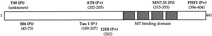
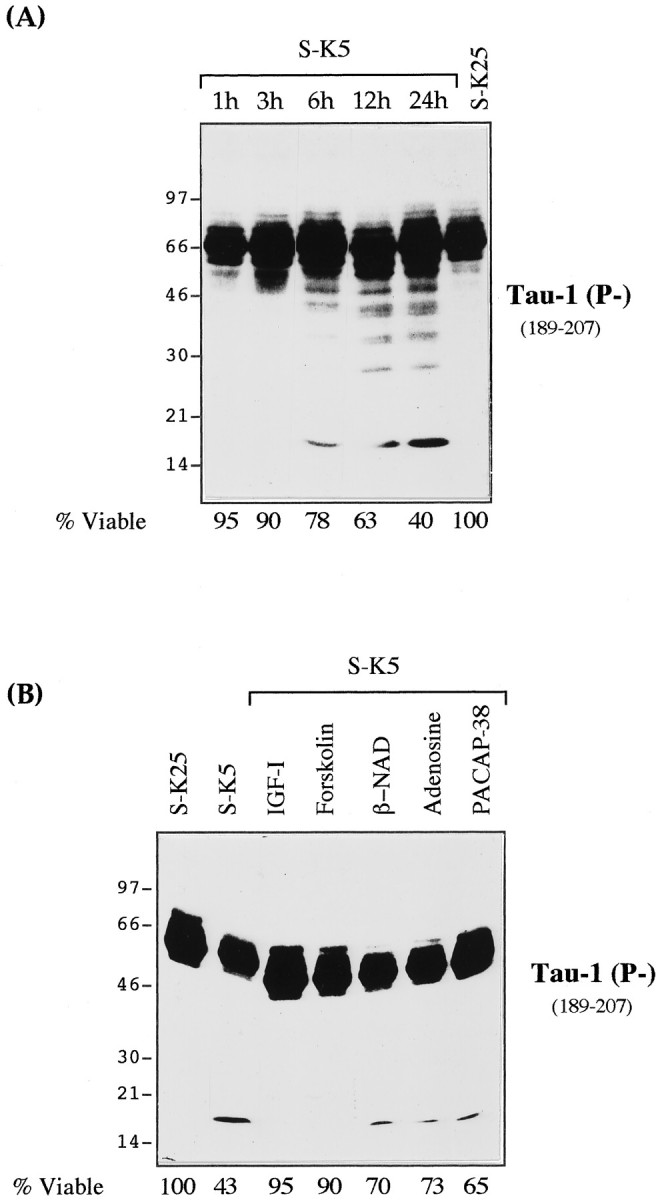
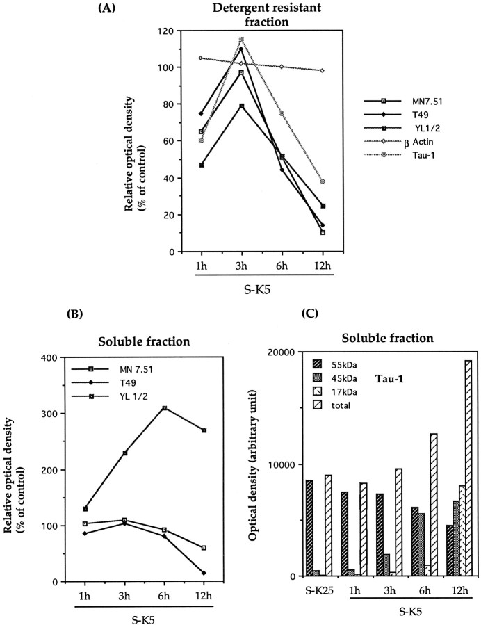
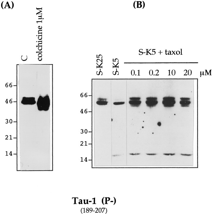
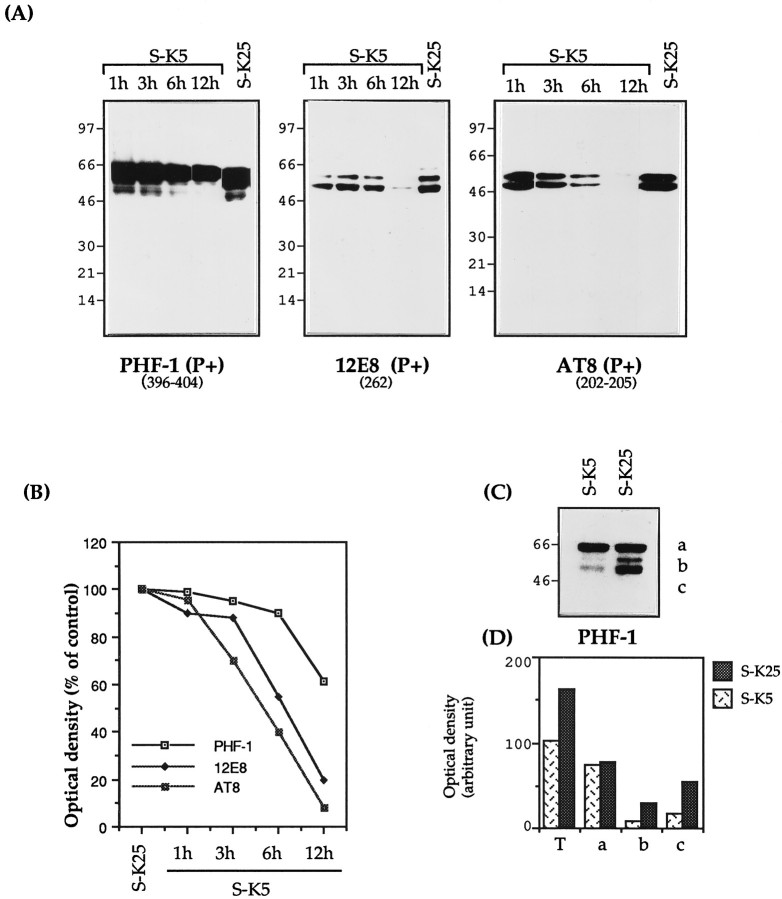
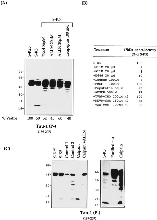
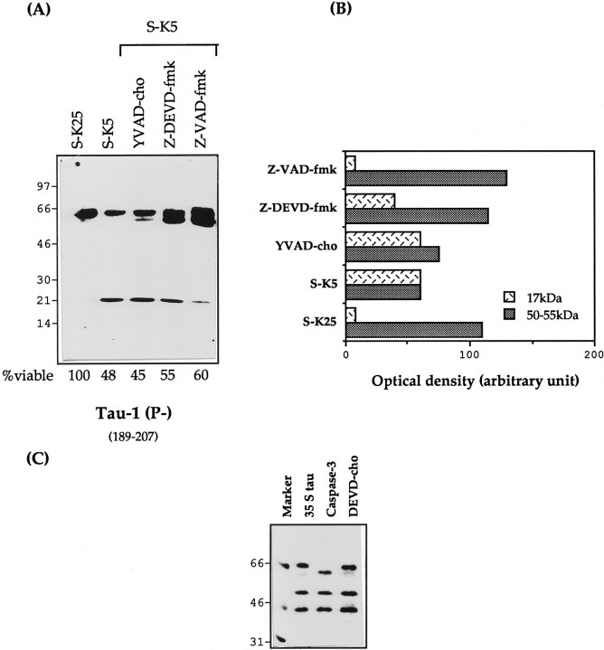
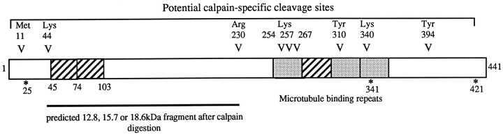
Cerebellar granule cells undergo apoptosis in culture after deprivation of potassium and serum. During this process we found that tau, a neuronal microtubule-associated protein that plays a key role in the maintenance of neuronal architecture, and the pathology of which correlates with intellectual decline in Alzheimer's disease, is cleaved. The final product of this cleavage is a soluble dephosphorylated tau fragment of 17 kDa that is unable to associate with microtubules and accumulates in the perikarya of dying cells. The appearance of this 17 kDa fragment is inhibited by both caspase and calpain inhibitors, suggesting that tau is an in vivo substrate for both of these proteases during apoptosis. Tau cleavage is correlated with disruption of the microtubule network, and experiments with colchicine and taxol show that this is likely to be a cause and not a consequence of tau cleavage. These data indicate that tau cleavage and change in phosphorylation are important early factors in the failure of the microtubule network that occurs during neuronal apoptosis. Furthermore, this study introduces new insights into the mechanism(s) that generate the truncated forms of tau present in Alzheimer's disease.


Figures

Fig. 1.
Fig. 1.
Tau antibodies used in this study. Schematic diagram of the longest tau isoform, showing the epitope locations of the antibodies used in this study. The antibodies used are indicated inbold, and the amino acid position of the epitopes is indicated in parentheses. The MT binding repeats arestippled. Antibodies 304, T49, and MN 7.51 recognize the primary tau sequence independently of phosphorylation status (indicated as Pi). 12E8, PHF-1, and AT8 recognize phosphorylated epitopes (P+), whereas Tau-1 binds to a dephosphorylated epitope (P−).
Fig. 2.
Fig. 2.
Specific and rapid cleavage of tau during the apoptosis of CGC induced by potassium and serum deprivation.A, CGCs were washed and maintained in high-potassium and serum-free medium (S-K25) for 24 hr or switched to K5 and serum-free BME (S-K5). At 1, 3, 6, 12, and 24 hr after the switch, 25 μg of total cellular protein was analyzed by Western blot using the mAb Tau-1 (see Fig. 1). For each time point, the corresponding viability was determined as described in Materials and Methods. B, Effect of various antiapoptotic agents on the generation of the Tau-1-recognized 17 kDa fragment. Granule cell neurons were maintained in S-K25 or subjected to S-K5 alone or in the presence of IGF-1 (25 ng/ml), forskolin (10 μm), nicotinamide adenine dinucleotide (β-NAD) (100 μm), adenosine (100 μm), or pituitary adenylate cyclase-activating polypeptide (PACAP)-38 (100 nm) for 16 hr. The corresponding viability for each lysate was determined as described in Material and Methods and reported below the figure.
Fig. 3.
Fig. 3.
The 17 kDa fragment is unable to bind to microtubules: disassembly of the microtubule network during apoptosis. Western blot analyses of cytoskeletal protein in cerebellar granule neurons undergoing apoptosis. Shown are Western blots of detergent-resistant (A, E) (30 μg) and soluble (F, I) fractions (20 μg) after Triton X-100 extraction as described in Material and Methods. Cerebellar granule neurons were cultured in the absence of serum and high potassium (S-K25), or the absence of serum and low potassium (S-K5). Western blot analysis was performed with the monoclonal antibodies Tau-1 (A,F) (specific for tau dephosphorylated at 189–207), MN 7.51 (B, G) (phosphorylation independent epitope in the third microtubule domain), T49 (C, H) (N-terminal epitope), YL 1/2 (D, I) (against α-tyrosinylated tubulin), and mAb to β-actin (E).
Fig. 4.
Fig. 4.
Densitometric analysis of Western blots reported in Figure 3. Tau, β-actin, and α-tyrosinylated tubulin immunoreactivity on detergent-resistant fractions (A) and soluble fractions (B) are expressed as percentages of control cells (S-K25 for 12 hr), where control cells have been given a value of 100. Results shown are representative of at least two experiments.C, Densitometric analysis of the different bands recognized by Tau-1 in Figure 3G. The absolute scanning values are given for 17, 45, and 55 kDa fragments.
Fig. 5.
Fig. 5.
Effect of colchicine and taxol on tau cleavage during apoptosis. A, Cultured cerebellar granule neurons were exposed to 1 μm colchicine for 24 hr or maintained in S+K25 [control (C)] or induced to undergo apoptosis by serum and potassium deprivation in the presence of taxol 0.1–20 μm for 12 hr. B, Total cellular proteins were extracted and analyzed by SDS-PAGE (7–15%) (15 μg of protein per lane in Fig. 4A and 25 μg per lane in Fig. 4B), electroblotted, and analyzed with mAb Tau-1.
Fig. 6.
Fig. 6.
Modulation of tau phosphorylation during apoptosis. A, Granule neurons were either maintained in high K+ medium (S-K25) for 12 hr or exposed to low K+ medium (S-K5) for various lengths of time. Total cellular proteins were extracted and analyzed by SDS-PAGE (7–15%) (15 μg of protein per lane), electroblotted, and analyzed with the mAbs PHF-1, 12E8, and AT8. B, Densitometric analysis of tau immunoreactivity expressed as percentages of control cells (untreated for 12 hr) where control cells have a value of 100, indicated for the different mAbs. C, PHF-1 immunoblotting showing the reduction of the 50–55 kDa isoforms and (D) their densitometric evaluation. Results are representative of at least two experiments. In Figure 5D, Tindicates the total PHF-1 immunoreactivity.
Fig. 7.
Fig. 7.
Immunofluorescence studies showing the effect of apoptosis on tau immunostaining. Shown is immunofluorescence of control cultures (A, A′, E) and cultures exposed to K+ and serum deprivation for 6 hr (C, C′, E′). Nuclei were stained with Hoechst 33258 (B, B′, D, D′, F, F′), and tau was immunostained with mAb MN 7.51 (A, C, E) or mAb Tau-1 (A′, C′, E′). Few or no apoptotic nuclei are seen in control cultures (B, B′). In D and D′, apoptotic nuclei, recognizable as those having a round shape and condensed or fragmented chromatin, are not immunostained or are minimally immunostained with MN 7.51 (C), whereas they are strongly stained with Tau-1 (C′). Big arrows indicate immunostaining with MN 7.51 and Tau-1 and their corresponding nuclei in the magnification picture (E, F, E′, F′). Scale bars, 7 μm.
Fig. 8.
Fig. 8.
Tau proteolysis profiles during apoptosis with different protease inhibitors. A, Total protein extracts (25 μg) of control cells (S-K25) and apoptotic cerebellar granule cells (S-K5) in the absence or presence of various calpain inhibitors: ALLN (20 μm), ALLM (20 μm), E64d (20 μm), and leupeptin (100 μm). All of the inhibitors were added 2 hr before and at the time of apoptosis induction. The lysates were resolved on a 7–15% gel and probed with mAb Tau-1. The corresponding viability for each lysate was determined as reported in Material and Methods. B, Densitometric values (% of S-K5) of the 17 kDa fragment after treatment with different protease inhibitors. C,In vitro cleavage of tau by calpain. S-K25, Control cells in 25 mm potassium; S-K5, cells induced to undergo apoptosis in 5 mm potassium. Control 1, Untreated cellular extracts incubated on ice. Control 2, Cellular extracts incubated with 5 mmCa2+ at 30°C for 10 min without calpain. Limited proteolysis of tau present in Control 2 occurs as a result of the activation of endogenous calpain by calcium. Calpain, Granule cell protein extract (20 μg of protein) or purified bovine tau treated with (0.5 μg) calpain at 30°C for 10 min;Calpain + ALLN, as calpain treated with ALLN 20 μm. The samples were analyzed on Western blot with mAb Tau-1.
Fig. 9.
Fig. 9.
Effect of caspase inhibitors on proteolysis of tau. A, Total protein extracts (25 μg) of control cells (S-K25) and apoptotic cerebellar granule cells (S-K5) in the presence or absence of various caspase inhibitors, YVAD-cho (100 μm), ZDEVD-fmk (150 μm), and ZVAD-fmk (100 μm), for 16 hr. All the inhibitors were added 2 hr before, and at the time of, apoptosis induction. The corresponding viability for each lysate was determined as reported in Materials and Methods. B, Densitometric analysis of the 17 kDa and the 50–55 kDa Tau-1 bands reported in A, expressed in densitometric units. Results are representative of at least two experiments. C, In vitro translated tau treated with bacterial extracts containing caspase-3. DEVD-cho is a specific inhibitor of caspase 3.
Fig. 10.
Fig. 10.
Map of tau with potential protease sites. The longest form of tau is indicated. V, Potential calpain sites. *, Potential caspase-3 sites. Hatched areas, Alternative spliced exons. Stippled areas, Microtubule binding repeats. Note that cleavage of tau at the N or C terminus by calpain or caspase would lead to production of a fragment of tau of similar molecular weight. Note that complete digestion of tau by calpain would lead to the production of 17 kDa fragment.

References

    1. Biernat J, Gustke N, Drewes G, Mandelkow EM, Mandelkow E. Phosphorylation at Ser 262 strongly reduces binding of tau to microtubules: distinction between PHF-like immunoreactivity and microtubule binding. Neuron. 1993;11:153–163. - PubMed
    1. Bonfoco E, Ceccatelli S, Manzo L, Nicotera L. Colchicine induces apoptosis in cerebellar granule cells. Exp Cell Res. 1995;218:189–200. - PubMed
    1. Brancolini C, Lazarevic D, Rodriguez J, Schneider C. Dismantling cell-cell contacts during apoptosis is coupled to caspase-dependent proteolytic cleavage of β-catenin. J Cell Biol. 1997;139:759–771. - PMC - PubMed
    1. Brandt R, Leger J, Lee G. Interaction of tau with the neural plasma membrana by tau’s amino-terminal projection domain. J Cell Biol. 1995;131:1327–1340. - PMC - PubMed
    1. Brion JP, Hanger DP, Bruce MT, Couck AM, Flament-Durand J, Anderton BH. Tau in Alzheimer neurofibrillary tangles. N- and C-terminal regions are differentially associated with paired helical filaments and the location of a putative abnormal phosphorylation site. Biochem J. 1991;273:127–133. - PMC - PubMed

Publication types

MeSH terms

LinkOut - more resources