. 1998 Sep 15;18(18):7138-51.
doi: 10.1523/JNEUROSCI.18-18-07138.1998.

Functional redundancy of FMRFamide-related peptides at the Drosophila larval neuromuscular junction

Affiliations

Functional redundancy of FMRFamide-related peptides at the Drosophila larval neuromuscular junction

R S Hewes et al. J Neurosci. .

Abstract

The Drosophila FMRFamide gene encodes multiple FMRFamide-related peptides. These peptides are expressed by neurosecretory cells and may be released into the blood to act as neurohormones. We analyzed the effects of eight of these peptides on nerve-stimulated contraction (twitch tension) of Drosophila larval body-wall muscles. Seven of the peptides strongly enhanced twitch tension, and one of the peptides was inactive. Their targets were distributed widely throughout the somatic musculature. The effects of one peptide, DPKQDFMRFamide, were unchanged after the onset of metamorphosis. The seven active peptides showed similar dose-response curves. Each had a threshold concentration near 1 nM, and the EC50 for each peptide was approximately 40 nM. At concentrations <0.1 microM, the responses to each of the seven excitatory peptides followed a time course that matched the fluctuations of the peptide concentration in the bath. At higher concentrations, twitch tension remained elevated for 5-10 min or more after wash-out of the peptide. When the peptides were presented as mixtures predicted by their stoichiometric ratios in the dFMRFamide propeptide, the effects were additive, and there were no detectable higher-order interactions among them. One peptide was tested and found to enhance synaptic transmission. At 0.1 microM, DPKQDFMRFamide increased the amplitude of the excitatory junctional current to 151% of baseline within 3 min. Together, these results indicate that the products of the Drosophila FMRFamide gene function as neurohormones to modulate the strength of contraction at the larval neuromuscular junction. In this role these seven peptides appear to be functionally redundant.


Figures
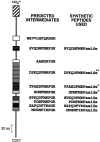
Fig. 1.
Fig. 1.
Predicted organization of the Drosophila melanogaster FMRFamide prepropeptide. Left, The deduced dFMRFamide gene product is shown by avertical line with boxes to indicate the locations of several neuropeptide sequences. Center, Possible peptide intermediates were selected on the basis of the fact that each sequence was at least 70% identical to a sequence in a similar position in the Drosophila virilis FMRFamidegene (Taghert and Schneider, 1990). Right, Each of the eight peptides synthesized for this study is bordered by consensus sites for proteolytic cleavage from the precursor (Devi, 1991), contains a putative amidation signal at the C terminus (Bleakman et al., 1988), and contains two or more residues matching the FMRF consensus sequence common to this family of peptides (Greenberg and Price, 1992). *Purified from Drosophila tissue byNichols (1992); **purified from Drosophila tissue byNambu et al. (1988) and Nichols (1992). Hatched box, Signal sequence.
Fig. 2.
Fig. 2.
Schematic diagram of the neuromuscular preparation. A, After removal of the gut and CNS, the anterior of each larva was pinned to the recording dish while the posterior was tethered to the arm of a calibrated strain gauge. Stimulation of an abdominal nerve with a suction electrode (2–6 V, 25–35 msec, 1 Hz) elicited contractions in the muscles of one abdominal half-segment (black muscles). Occasionally, the nerve stimulus also elicited contractions in muscles of adjacent segments (nearest of the muscles shown ingray). B, Changes in the bath dye concentration/applied dye concentration as a function of time. Three successive trials were performed. In trials 1 and3, the dye (fast green) was applied to the recording chamber at time = 0 min. In trial 2, 1 ml of saline was used as a wash. The perfusion rate in trials 1 and2 (1.0 ml/min) was equal to the fastest rate used in the contraction assays. The perfusion rate in trial 3 (0.6 ml/min) was close to the slowest rate used in the assays.
Fig. 3.
Fig. 3.
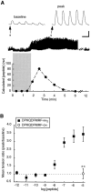
DPKQDFMRFamide, the most abundant sequence in theDrosophila FMRFamide gene, enhances nerve-stimulated contractions of abdominal muscles. A, Recording of muscle contractions in a wandering third instar larva; 100 nm DPKQDFMRFamide was applied to the preparation during the period indicated by the shaded rectangle at a perfusion rate of 0.6 ml/min. Periods indicated by the black squares are shown at an expanded time scaleabove. The calculated changes in bath peptide concentration (derived from trial 3, Fig.2B) are plotted below at the same time scale. Before peptide application (baseline), each stimulation of the nerve elicited weak contractions in 5–10 of the 30 muscle fibers in one abdominal half-segment. At the time of peak response (peak), peak tension was increased by 456%. This increase in tension was accompanied by an increase in the number of strongly contracting fibers. B, Mean tension ratio ± SEM as a function of DPKQDFMRFamide (n = 7) or DPKQDFMRF-OH (free acid; n = 5) concentration in third instar larvae. Tension ratio = peak tension (the mean amplitude of five contractions 230 sec after the beginning of a given peptide application)/baseline tension (the mean amplitude of 10 contractions before peptide application—see Materials and Methods). **p < 0.01 (Student’s t test). Calibration: Top traces, 0.2 nN, 1 sec; bottom trace, 0.26 nN, 40 sec.
Fig. 4.
Fig. 4.
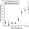
The effects of DPKQDFMRFamide on muscle contraction do not change during progression from the feeding to the wandering larval stage. Dose–response curves (mean ± SEM) for DPKQDFMRFamide are plotted separately for feeding-stage (n ≥ 5) and wandering-stage (n= 7) larvae (wandering-stage data the same as shown in Fig.3B).
Fig. 5.
Fig. 5.
The excitatory effects of each of the seven active peptides are slow to develop and are long-lasting; the time courses of the responses are similar. Peptides (each at 10-7m) were applied to the preparations during the periods indicated by the horizontal bars. The illustrated differences in kinetics reflect the differences among different preparations rather than consistent differences among the effects of the different peptides. Baseline shifts such as those indicated (small arrows) are attributable to subtle changes in the bath volume and have no detectable effect on contraction amplitudes. Slower baseline shifts in some preparations accompanied peptide-mediated increases in nerve-stimulated contractions (open arrows), particularly at higher peptide concentrations. These shifts are attributable to a decrease in the relaxation rate, which causes summation (an apparent increase in baseline tension). In control preparations in which the frequency of nerve stimulation is varied, this summation does not alter the amplitudes of individual contractions. Calibration: 200 pN, 100 sec.
Fig. 6.
Fig. 6.
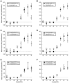
Mean tension ratio ± SEM as a function of peptide concentration for each of the predicted excitatory products of the FMRFamide gene (wandering third instar larvae). For clarity, the results were plotted in six sets, and the dose–response curve for DPKQDFMRFamide (see Fig. 3B,n = 7) was included in each set for reference. However, at each concentration a single ANOVA was performed on the complete data set, containing the results for all seven active peptides. Individual means were compared by post hoctesting subsequent to the ANOVA. *p < 0.05; **p < 0.01 (Spjotvoll–Stoline) [one-way ANOVA; 10-11m,F(6,55) = 3.164, p = 0.0097; 10-10m,F(6,55) = 4.810, p = 0.0005; 10-9m,F(6,55) = 7.896, p = 0.0001; 10-6m,F(6,55) = 3.319, p = 0.0073; 10-5m,F(6,55) = 7.353, p = 0.0001]. n = 10, SPKQDFMRFamide, SDNFMRFamide, and TPAEDFMRFamide; n ≥ 9, PDNFMRFamide;n ≥ 7, SVQDNFMHFamide; n ≥ 8, MDSNFIRFamide.
Fig. 7.
Fig. 7.
SAPQDFVRSamide did not enhance twitch tension. The mean tension ratio ± SEM (n = 6) at each concentration was measured at 230 sec (the average time to peak for DPKQDFMRFamide). The data for DPKQDFMRFamide are taken from Figure3B (n = 7). *p< 0.05; **p < 0.01; ***p < 0.001 [one-way ANOVA: 10-12m,F(1,11) = 6.329, p = 0.0287; 10-8m,F(1,11) = 9.444, p = 0.0106; 10-7m,F(1,11) = 72.601, p = 0.0001; 10-6m,F(1,11) = 37.506, p = 0.0001; 10-5m,F(1,11) = 72.170, p = 0.0001].
Fig. 8.
Fig. 8.
The effects of stoichiometric molar ratios of dFMRFamide-related peptides are additive. A–C, Mean tension ratio ± SEM is shown as a function of total peptide concentration, using a peptide mixture (“mix 3”) containing DPKQDFMRFamide, TPAEDFMRFamide, and SDNFMRFamide in a 5:2:1 molar ratio (“mix 3,” n = 10; DPKQDFMRFamide,n = 7). The predicted curve (solid line) was generated by averaging the results for DPKQDFMRFamide, TPAEDFMRFamide, and SDNFMRFamide in the same 5:2:1 ratio (data from the same recordings as in Fig. 6). Peak twitch tension measurements for “mix 3,” DPKQDFMRFamide, and the predicted curve were taken at 130 sec (A), 180 sec (B), or 230 sec (C) after the onset of peptide application. D, Mean tension ratio ± SEM measured at 150, 190, and 260 sec after the onset of peptide application. The six treatments were used in a double-blind experiment in a randomized order (n = 14 for all treatments). The differences in tension measured at 150, 190, and 260 sec with the blank control were not statistically significant (one-way ANOVA). DPK −8, 10-8mDPKQDFMRFamide; DPK −9, 10-9m DPKQDFMRFamide; mix 3 −9, 10-9m, 3 peptide mix in 5:2:1 ratio;mix 8 −9, 10-9m, 8 peptide mix in 5:2:1:1:1:1:1:1 ratio; TPA −9, 10-9m TPAEDFMRFamide;blank, saline control. *p < 0.05; **p < 0.01 (Spjotvoll–Stoline) [data inD, one-way ANOVA: 190 sec,F(5,78) = 5.091, p = 0.0004; 260 sec, F(5,78) = 3.637,p = 0.0052].
Fig. 9.
Fig. 9.
A Drosophila myosuppressin, TDVDHVFLRFamide, enhances muscle contraction, but it is active only at micromolar concentrations. Mean tension ratio ± SEM is shown as a function of TDVDHVFLRFamide concentration in wandering third instar larvae (n = 6). The mean time-to-peak tension was 150.8 ± 7.4 (n = 18). The data for DPKQDFMRFamide are taken from Figure 3B(n = 7). *p < 0.05; **p < 0.001; ***p < 0.001 [one-way ANOVA: 10-8m,F(1,11) = 7.428, p = 0.0197; 10-7m,F(1,11) = 53.424, p = 0.0001; 10-6m,F(1,11) = 19.745, p = 0.0010; 10-5m,F(1,11) = 9.318, p = 0.0110].
Fig. 10.
Fig. 10.
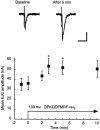
DPKQDFMRFamide enhances synaptic transmission at the larval neuromuscular junction. Excitatory junctional currents (EJCs) were evoked in muscle fiber 6 by electrical stimulation of the abdominal nerve. Representative EJCs (preceded by the stimulus artifact) are shown above. The mean EJC amplitude ± SEM as a function of time after the application of 100 nm DPKQDFMRFamide is plotted below(n = 4–6 at each point). *p < 0.05 (Student’s t test, compared with the baseline mean). Calibration: 20 nA, 20 msec.

Similar articles

Cited by

References

    1. Anderson MS, Halpern ME, Keshishian H. Identification of the neuropeptide transmitter proctolin in Drosophila larvae: characterization of muscle fiber-specific neuromuscular endings. J Neurosci. 1988;8:242–255. - PMC - PubMed
    1. Bainbridge SP, Bownes M. Staging the metamorphosis of Drosophila melanogaster. J Embryol Exp Morphol. 1981;66:57–80. - PubMed
    1. Bendena WG, Garside CS, Yu CG, Tobe SS. Allatostatins: diversity in structure and function of an insect neuropeptide family. Ann NY Acad Sci. 1997;814:53–66. - PubMed
    1. Benjamin PR, Burke JF. Alternative mRNA splicing of the FMRFamide gene and its role in neuropeptidergic signalling in a defined neural network. BioEssays. 1994;16:335–342. - PubMed
    1. Bleakman A, Bradbury AF, Darby NJ, Maruthainar K, Smyth DG. Processing reactions in the later stages of hormone activation. Biochimie. 1988;70:3–10. - PubMed

Publication types

MeSH terms

LinkOut - more resources