. 1998 Sep 15;18(18):7152-9.
doi: 10.1523/JNEUROSCI.18-18-07152.1998.

Central P2X4 and P2X6 channel subunits coassemble into a novel heteromeric ATP receptor

Affiliations

Central P2X4 and P2X6 channel subunits coassemble into a novel heteromeric ATP receptor

K T Lê et al. J Neurosci. .

Abstract

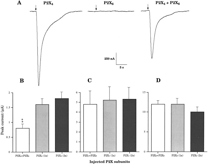
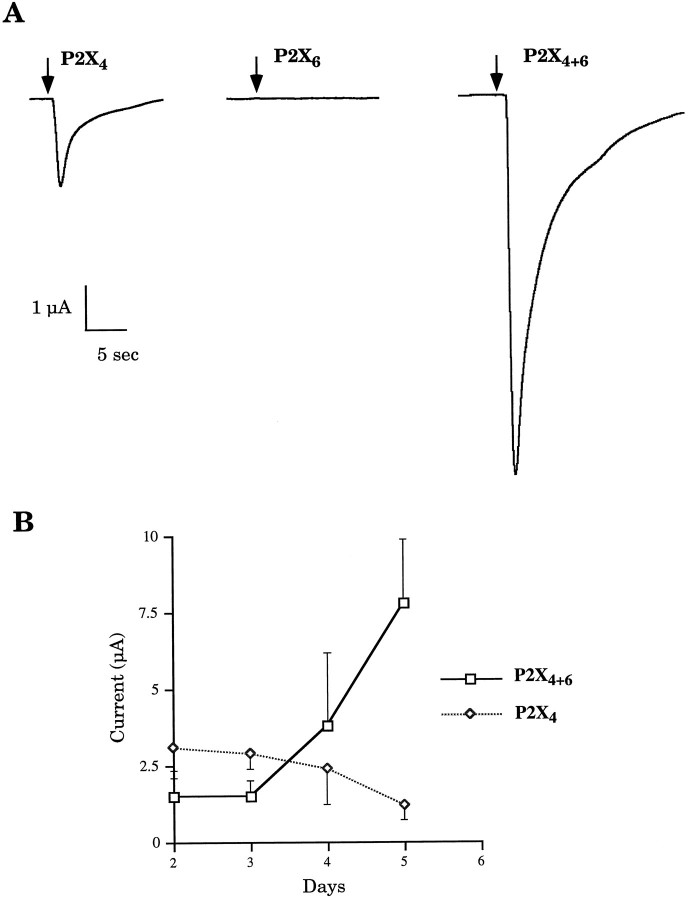
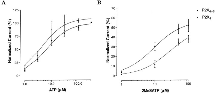
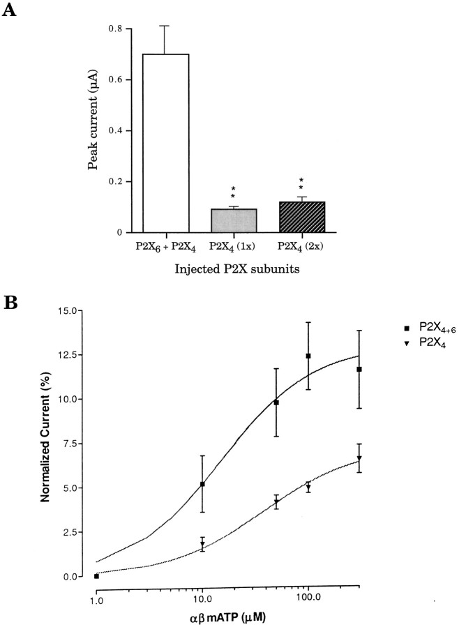
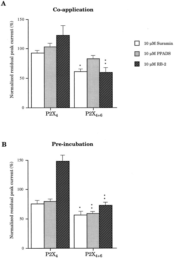
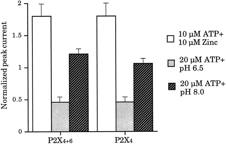
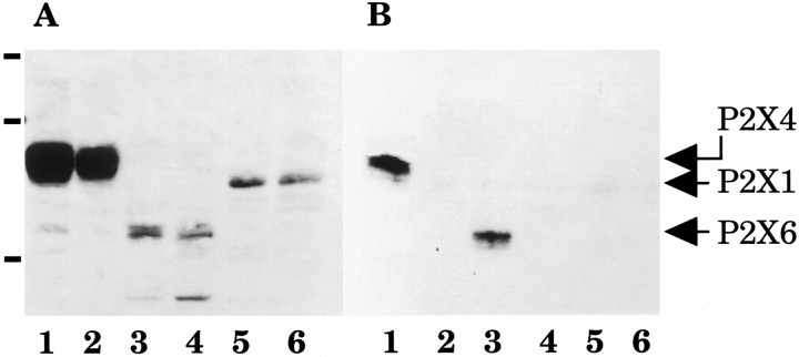
Ionotropic ATP receptors are widely expressed in mammalian CNS. Despite extensive functional characterization of neuronal homomeric P2X receptors in heterologous expression systems, the subunit composition of native central P2X ATP-gated channels remains to be elucidated. P2X4 and P2X6 are major central subunits with highly overlapping mRNA distribution at both regional and cellular levels. When expressed alone in Xenopus oocytes, P2X6 subunits do not assemble into surface receptors responsive to ATP applications. On the other hand, P2X4 subunits assemble into bona fide ATP-gated channels, slowly desensitizing and weakly sensitive to the partial agonist alpha,beta-methylene ATP and to noncompetitive antagonists suramin and pyridoxal-5-phosphate-6-azophenyl-2',4'-disulfonic acid. We demonstrate here that the coexpression of P2X4 and P2X6 subunits in Xenopus oocytes leads to the generation of a novel pharmacological phenotype of ionotropic ATP receptors. Heteromeric P2X4+6 receptors are activated by low-micromolar alpha, beta-methylene ATP (EC50 = 12 microM) and are blocked by suramin and by Reactive Blue 2, which has the property, at low concentrations, to potentiate homomeric P2X4 receptors. The assembly of P2X4 with P2X6 subunits results from subunit-dependent interactions, as shown by their specific copurification from HEK-293 cells transiently transfected with various epitope-tagged P2X channel subunits. Our data strongly suggest that the numerous cases of neuronal colocalizations of P2X4 and P2X6 subunits observed in mammalian CNS reflect the native expression of heteromeric P2X4+6 channels with unique functional properties.


Figures

Fig. 1.
Fig. 1.
Representative heteromeric P2X4+6channel current phenotype at day 3. A, ATP-induced currents after heterologous expression of P2X4, P2X6, and P2X4 + P2X6 (1:1 molar ratio) subunits recorded 3 d after corresponding cDNA nuclear microinjections in Xenopus oocytes.Arrows indicate beginnings of ATP applications (10 sec).B, P2X4-dependent functional impact of P2X6 on ATP-induced response (P2X4 expressed alone; 1x, 5 ng of cDNA; 2x, 10 ng).C, P2X1 receptor (P2X1;1x, 5 ng; 2x, 10 ng) functional expression is unaffected by coexpressed P2X6 subunits.D, P2X2-mediated (P2X2;1x, 5 ng; 2x, 10 ng) ATP-induced peak current amplitudes are unchanged in the presence of P2X6subunits. (Averages ± SEM from 3 to 15 oocytes in 2–4 independent experiments; double asterisks denote significant difference; p < 0.01).
Fig. 2.
Fig. 2.
A, Potentiation of ATP response represented by P2X4+6 channel current phenotype at day 5.Arrows indicate beginnings of ATP applications (10 sec).B, Time course of heteromeric P2X4+6expression. Kinetics of appearance of functional ATP receptors on plasma membranes is strikingly different in oocytes coinjected with P2X4 and P2X6 subunits compared with those injected with P2X4 subunits (averages ± SEM from 3 to 15 oocytes in 2–8 independent experiments).
Fig. 3.
Fig. 3.
Sensitivity of P2X4+6 receptors to the agonists ATP and 2MeSATP. A, Similar ATP dose–response profile between heteromeric P2X4+6 channels and homomeric P2X4 receptors in Xenopus oocytes.B, Heteromeric P2X4+6 receptors showed increased sensitivity to 2MeSATP compared with P2X4receptors. Values are normalized to the response to 300 μm ATP (averages ± SEM from 3 to 7 oocytes per point in 2 independent experiments).
Fig. 4.
Fig. 4.
Sensitivity of P2X4+6 receptors to the agonist αβmATP. A, Differential αβmATP responsiveness measured in peak current amplitudes betweenXenopus oocytes expressing either P2X4+6channels or P2X4 at day 3 after injection; see Figure1B for comparison of ATP-induced peak currents.B, Normalized dose–response curves of P2X4+6 and P2X4 receptor species for αβmATP. Values are normalized to the response to 100 μm ATP; double asterisks denote significant difference; p < 0.01 (averages ± SEM from 5 to 8 oocytes per point in 2 independent experiments).
Fig. 5.
Fig. 5.
Sensitivity of P2X4+6 to P2X antagonists. Suramin, PPADS, and RB-2 were tested for their blocking properties on heteromeric P2X4+6 channels and on homomeric P2X4 receptors. Antagonists were coapplied with ATP (A) or preincubated before coapplication (B). Note that P2X4+6 receptors are inhibited, whereas P2X4 receptors are potentiated by 10 μm RB-2. Values are normalized to the response to ATP only (averages ± SEM from 5 oocytes per experiment;single and double asterisks denote significant difference; p < 0.05 andp < 0.01, respectively).
Fig. 6.
Fig. 6.
Sensitivity of P2X4+6 to the extracellular cofactors zinc ions and pH. Extracellular Zn2+, pH 6.5 and 8.0, coapplied with ATP, did not allow differentiation between P2X4+6 and P2X4receptors. Values are normalized to the response to ATP only (averages ± SEM from 4 oocytes per experiment).
Fig. 7.
Fig. 7.
Subunit specificity of P2X4 and P2X6 heteropolymerization. A, Immunoblot of Flag-tagged P2X1, P2X4, and P2X6 subunits probed with anti-Flag M2 monoclonal antibodies in total membrane proteins from transiently transfected HEK-293A cells. B, From the same samples, immunoblot of Flag-tagged P2X1, P2X4, and P2X6 subunits probed with M2 antibodies after copurification through P2X4-(His)6 subunits. Molecular weight markers (in A): 104, 82, and 48 kDa. Cotransfections: lane 1, P2X4-(His)6 + P2X4-Flag;lane 2, P2X4-wt + P2X4-Flag;lane 3, P2X4-(His)6 + P2X6-Flag; lane 4, P2X4-wt + P2X6-Flag; lane 5, P2X4-(His)6 + P2X1-Flag;lane 6, P2X4-wt + P2X1-Flag.

Similar articles

Cited by

References

    1. Balcar VJ, Li Y, Killinger S, Bennett MR. Autoradiography of P2X ATP receptors in the rat brain. Br J Pharmacol. 1995;115:302–306. - PMC - PubMed
    1. Bo X, Burnstock G. Distribution of [3H]α,β-methylene ATP binding sites in rat brain and spinal cord. NeuroReport. 1994;5:1601–1604. - PubMed
    1. Bo X, Zhang Y, Nassar M, Burnstock G, Schoepfer R. A P2X purinoceptor cDNA conferring a novel pharmacological profile. FEBS Lett. 1995;375:129–133. - PubMed
    1. Brake AJ, Wagenbach MJ, Julius D. New structural motif for ligand-gated ion channels defined by an ionotropic ATP receptor. Nature. 1994;371:519–523. - PubMed
    1. Buell G, Collo G, Rassendren F. P2X receptors: an emerging channel family. Eur J Neurosci. 1996;8:2221–2228. - PubMed

Publication types

MeSH terms

LinkOut - more resources