. 1998 Sep 15;18(18):7296-305.
doi: 10.1523/JNEUROSCI.18-18-07296.1998.

Regional selective neuronal degeneration after protein phosphatase inhibition in hippocampal slice cultures: evidence for a MAP kinase-dependent mechanism

Affiliations

Regional selective neuronal degeneration after protein phosphatase inhibition in hippocampal slice cultures: evidence for a MAP kinase-dependent mechanism

E Rundén et al. J Neurosci. .

Abstract

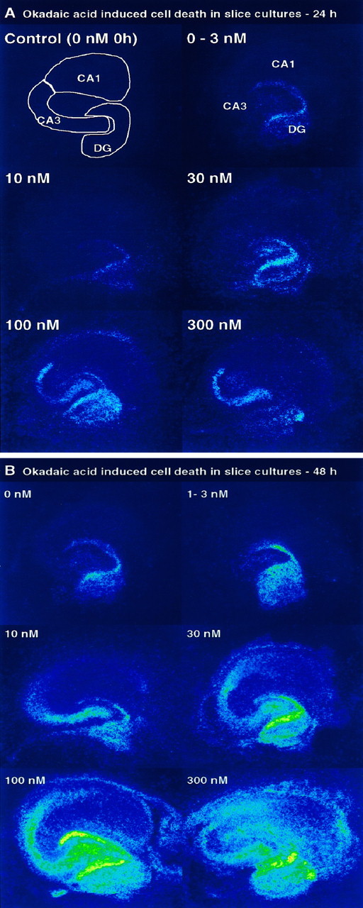
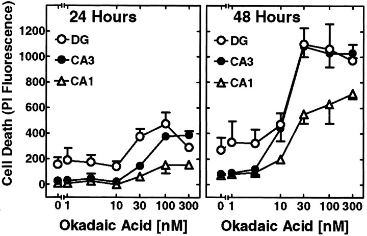
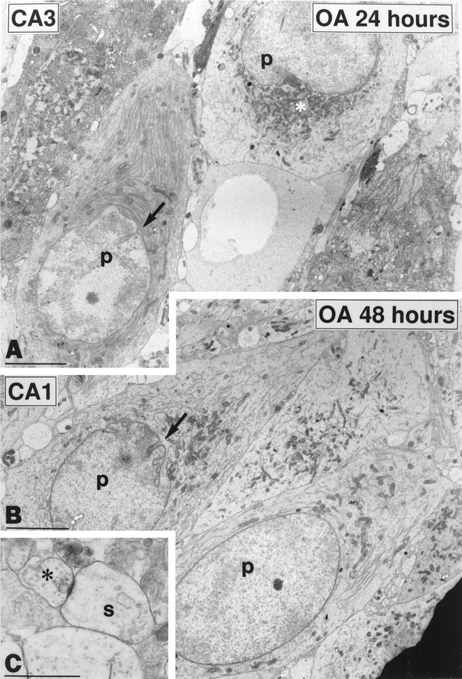
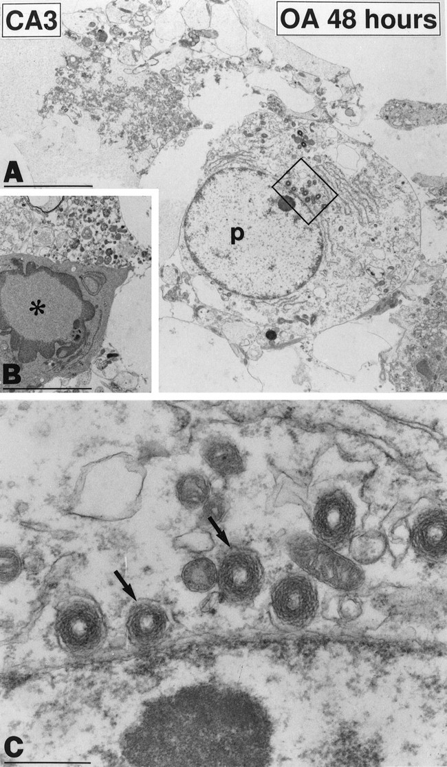
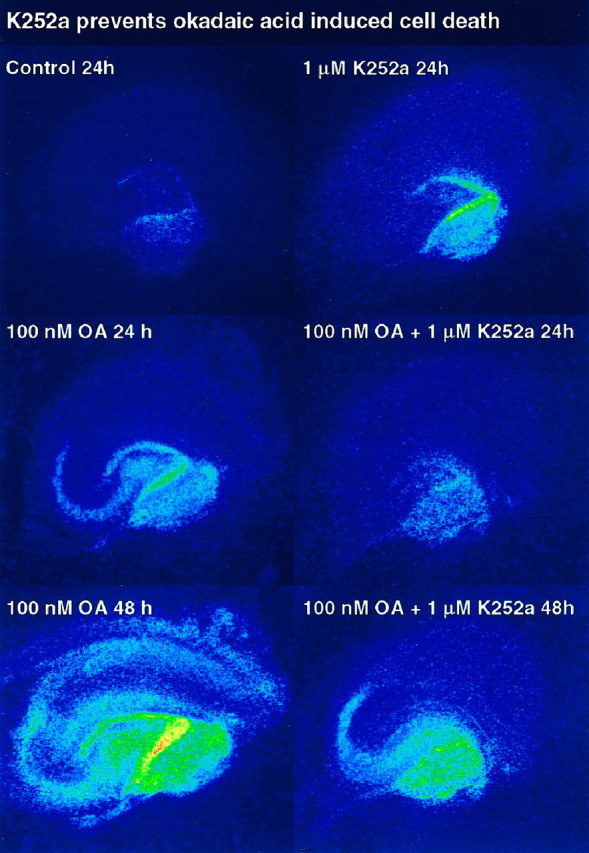
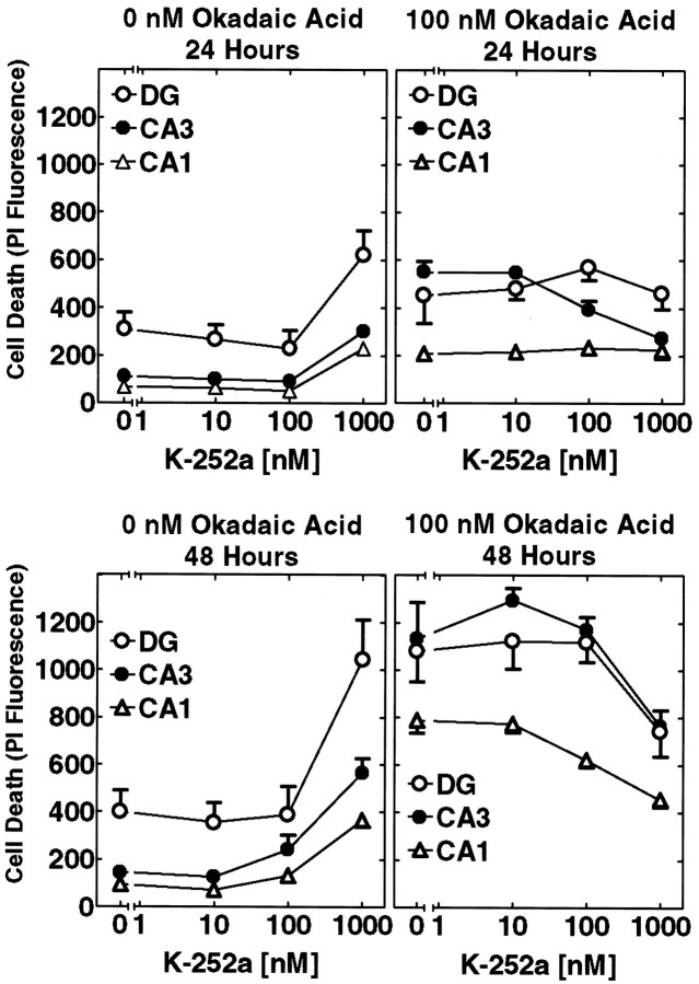
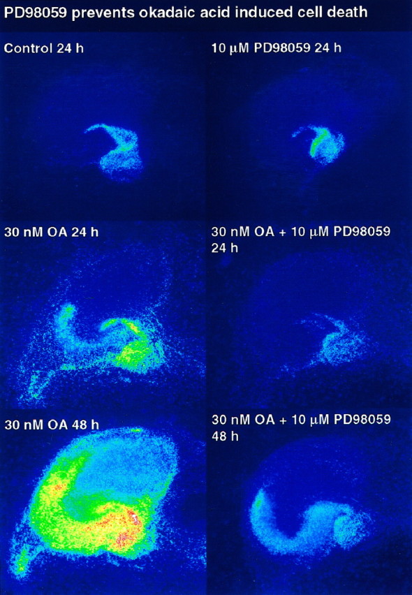
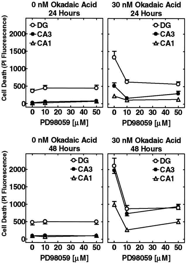
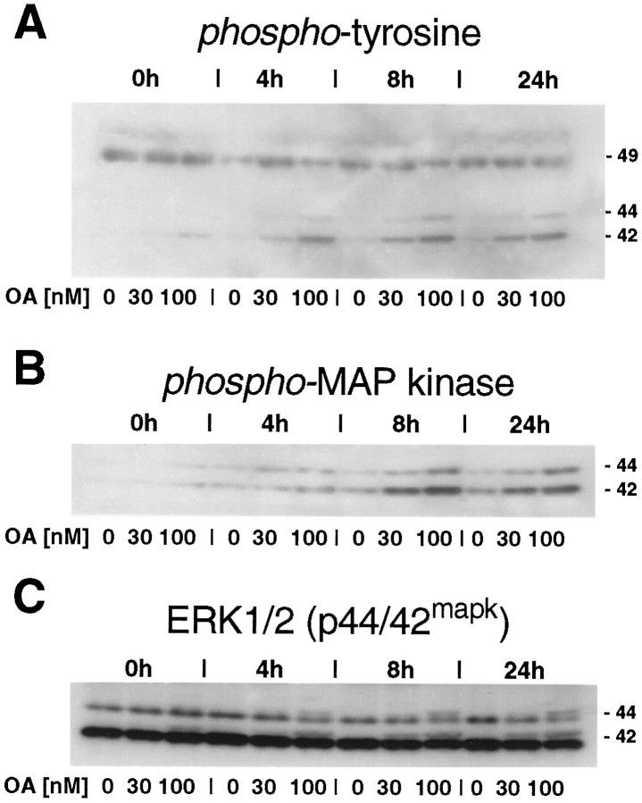
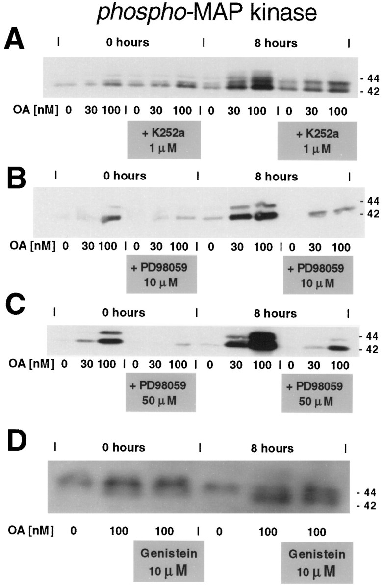
The regional selectivity and mechanisms underlying the toxicity of the serine/threonine protein phosphatase inhibitor okadaic acid (OA) were investigated in hippocampal slice cultures. Image analysis of propidium iodide-labeled cultures revealed that okadaic acid caused a dose- and time-dependent injury to hippocampal neurons. Pyramidal cells in the CA3 region and granule cells in the dentate gyrus were much more sensitive to okadaic acid than the pyramidal cells in the CA1 region. Electron microscopy revealed ultrastructural changes in the pyramidal cells that were not consistent with an apoptotic process. Treatment with okadaic acid led to a rapid and sustained tyrosine phosphorylation of the mitogen-activated protein kinases ERK1 and ERK2 (p44/42(mapk)). The phosphorylation was markedly reduced after treatment of the cultures with the microbial alkaloid K-252a (a nonselective protein kinase inhibitor) or the MAP kinase kinase (MEK1/2) inhibitor PD98059. K-252a and PD98059 also ameliorated the okadaic acid-induced cell death. Inhibitors of protein kinase C, Ca2+/calmodulin-dependent protein kinase II, or tyrosine kinase were ineffective. These results indicate that sustained activation of the MAP kinase pathway, as seen after e.g., ischemia, may selectively harm specific subsets of neurons. The susceptibility to MAP kinase activation of the CA3 pyramidal cells and dentate granule cells may provide insight into the observed relationship between cerebral ischemia and dementia in Alzheimer's disease.


Figures

Fig. 1.
Fig. 1.
Okadaic acid causes selective neuronal degeneration. A, False color-coded images of PI-labeled hippocampal slice cultures. Images were obtained at the start of the experiment and at 24 and 48 hr. The top left image is from a control culture at the start of the experiment (0 nmokadaic acid, 0 hr). It shows the outline of the different subregions of the slice cultures [CA1, CA3, and dentate gyrus (DG)]. The other images are from cultures incubated for 24 hr after treatment with okadaic acid (concentrations indicated). Cultures (13 DIV) were incubated in serum-free medium with PI and OA (0–300 nm). PI fluorescence indicates cell death.B, Similar images as in A obtained at 48 hr.
Fig. 2.
Fig. 2.
Dose–response curves of OA-induced cell death. The experimental conditions were as in Figure 1. PI fluorescence intensities were measured from the outlined regions illustrated in Figure 1A (DG, CA3, andCA1) after incubation with OA (0–300 nm). Student’s t test showed a significant increase in cell death in all regions at OA concentrations >10 nm(p < 0.05, Student’s ttest). Error bars indicate SEM; n = 5.
Fig. 3.
Fig. 3.
Okadaic acid-induced cell damage. Ultrastructural changes in hippocampal pyramidal cells. Cultures (13 DIV) were treated with 100 nm OA in serum-free medium for 24 hr, after which they were fixed by immersion in 2.5% glutaraldehyde and 1% paraformaldehyde and prepared for flat-embedding in epoxy resin. Ultrathin sections were studied with a Philips CM10 transmission electron microscope. A, The ultrastructural changes seen in the pyramidal cells (p) of the CA3 region at 24 hr involved slight nuclear indentations (arrow), aggregations of mitochondria around the nuclei (asterisk), and an increased amount of endoplasmic reticulum. Many cells also exhibited gross damage. Scale bar, 5 μm.B, At 48 hr, similar but less extensive changes were seen in pyramidal cells (p) in the CA1 region. Scale bar, 5 μm. C, Synaptic contacts in the CA1 region at 48 hr exhibit swollen dendritic spines (s) and nerve terminals (asterisk) with dispersed vesicles. Scale bar, 1 μm.
Fig. 4.
Fig. 4.
Okadaic acid-induced cell damage. Electron microscopic images from the CA3 region after incubation with 100 nm OA for 48 hr. A, Pyramidal cells (p) exhibited extensive damage with disrupted cytoplasm. The nuclei were usually morphologically intact. Scale bar, 5 μm. B, Glial cell (asterisk) in the CA3 region with morphological features reminiscent of apoptosis, i.e., multiple nuclear indentations and condensation of the chromatin. Scale bar, 5 μm. C, Enlarged image of framed area inA showing multiple spherical cytoplasmic bodies (arrows) composed of concentric osmophilic lamellae, possibly degenerating mitochondria, surrounding the nucleus. Scale bar, 0.5 μm.
Fig. 5.
Fig. 5.
K-252a protects against okadaic acid-induced cell death. False color-coded images of propidium iodide-labeled slice cultures incubated for 24–48 hr. Cultures (13 DIV) were incubated in serum-free medium containing PI and the nonspecific kinase inhibitor K-252a (0–1000 nm). Images were obtained with or without addition of 100 nm OA and at 24 and 48 hr.
Fig. 6.
Fig. 6.
K-252a protects against okadaic acid-induced cell death. Dose–response curves illustrate both the toxic effects and amelioration of OA-induced cell death by K-252a. Experimental conditions were as in Figure 5. Significant reduction in OA-induced cell death was seen in the CA3 region at 24 hr in cultures treated with 100–1000 nm K-252a and in all regions at 48 hr (p < 0.05, SEM; n = 5; Student’s t test).
Fig. 7.
Fig. 7.
PD98059 protects against okadaic acid-induced cell death. False color-coded images of propidium iodide-labeled slice cultures incubated for 24–48. Cultures (13 DIV) were preincubated for 1 hr in serum-free medium containing PI and the MEK1/2 inhibitor PD98059 (10 μm). Images were obtained after addition of OA (0 or 30 nm) and at 24 and 48 hr.
Fig. 8.
Fig. 8.
PD98059 protects against okadaic acid-induced cell death. Dose–response curves illustrate the amelioration of OA-induced cell death by PD98059. Experimental conditions were as in Figure 7. Significant reduction in OA-induced cell death was seen in all regions at 24 and 48 hr in cultures treated with 10–50 μmPD98059 (p < 0.05, SEM;n = 5; Student’s t test).
Fig. 9.
Fig. 9.
Okadaic acid-induced hyperphosphorylation of MAP kinase. Cultures (13 DIV) were incubated in serum-free medium. After addition of OA (0–100 nm) or vehicle (DMSO) to all the cultures, some were removed into Eppendorf tubes and frozen in liquid nitrogen. As much as 15 min may have passed from the beginning of the addition of OA until the first group of cultures was frozen. Lanes with labeled tissue from these cultures are labeled 0h on the blots. Other cultures were removed at 4 hr, 8 hr, and 24 hr. Each tissue sample was pooled from five cultures and subjected to SDS-PAGE.A, Western blot of OA-treated cultures labeled with an antibody against phosphotyrosine. B, Same blot as inA but with an antibody recognizing the phosphorylated form of MAP kinase (phospho-MAP kinase).C, Same blot as in A but with an antibody against unphosphorylated MAP kinase (ERK1/2–p44/42mapk). Note mobility shift at high doses of OA.
Fig. 10.
Fig. 10.
Reduction in MAP kinase phosphorylation with the protein kinase inhibitor K-252a and with the MEK1/2 inhibitor PD98059. Cultures (13 DIV) were incubated in serum-free medium containing the bacterial alkaloid K-252a (1 μm) (A) or the MEK1/2 inhibitor PD98059 (10 and 50 μm) (B, C). After addition of OA (0–100 nm), some cultures were removed and frozen. This procedure took ∼15 min (labeled 0 hours on the blot). Others were removed after 8 hr. Each tissue sample was pooled from five cultures and subjected to SDS-PAGE. D, Cultures were treated essentially as in Figure 9 but with the addition of the tyrosine kinase inhibitor genistein (10 μm).

Similar articles

Cited by

References

    1. Alessi DR, Cuenda A, Cohen P, Dudley DT, Saltiel AR. PD 098059 is a specific inhibitor of the activation of mitogen-activated protein kinase kinase in vitro and in vivo. J Biol Chem. 1995;270:27489–27494. - PubMed
    1. Ankarcrona M, Dypbukt JM, Orrenius S, Nicotera P. Calcineurin and mitochondrial function in glutamate-induced neuronal cell death. FEBS Lett. 1996;394:321–324. - PubMed
    1. Arendt T, Holzer M, Grossmann A, Zedlick D, Bruckner MK. Increased expression and subcellular translocation of the mitogen-activated protein kinase kinase and mitogen-activated protein kinase in Alzheimer’s disease. Neuroscience. 1995;68:5–18. - PubMed
    1. Benito A, Lerga A, Silva M, Leon J, Fernandez-Luna JL. Apoptosis of human myeloid leukemia cells induced by an inhibitor of protein phosphatases (okadaic acid) is prevented by Bcl-2 and Bcl-XL. Leukemia. 1997;11:940–944. - PubMed
    1. Billingsley ML, Kincaid RL. Regulated phosphorylation and dephosphorylation of tau protein: effects on microtubule interaction, intracellular trafficking and neurodegeneration. Biochem J. 1997;323:577–591. - PMC - PubMed

Publication types

MeSH terms

Substances

LinkOut - more resources