. 1998 Sep 15;18(18):7351-60.
doi: 10.1523/JNEUROSCI.18-18-07351.1998.

The survival-promoting effect of glial cell line-derived neurotrophic factor on axotomized corticospinal neurons in vivo is mediated by an endogenous brain-derived neurotrophic factor mechanism

Affiliations

The survival-promoting effect of glial cell line-derived neurotrophic factor on axotomized corticospinal neurons in vivo is mediated by an endogenous brain-derived neurotrophic factor mechanism

K M Giehl et al. J Neurosci. .

Abstract

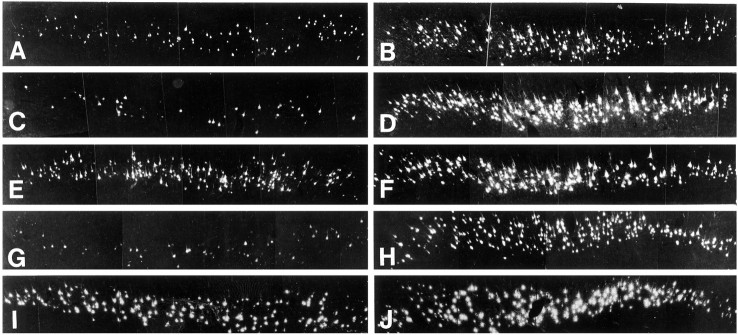
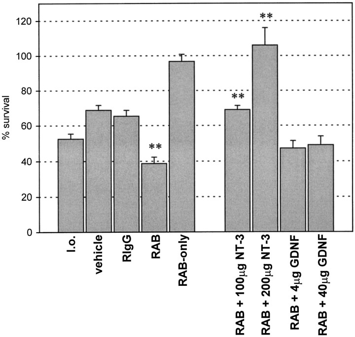
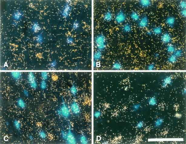
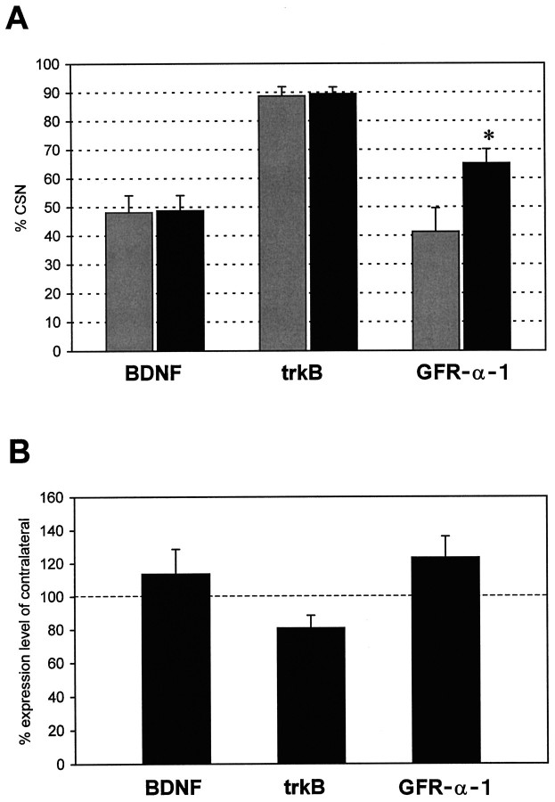
Autocrine trophic functions of brain-derived neurotrophic factor (BDNF) have been proposed for many central neurons because this neurotrophin displays striking colocalization with its receptor trkB within the CNS. In the cortex, the distribution patterns of BDNF and trkB expression are almost identical. Corticospinal neurons (CSNs) are a major cortical long-distance projecting system. They are localized in layer V of the somatosensory cortex, and their axons project into the spinal cord where they contribute to the innervation of spinal motoneurons. We have shown recently that adult CSNs express trkB mRNA and are rescued from axotomy-induced death by BDNF treatment. Half of the axotomized CSNs survived without BDNF infusions. These findings raise the possibility that endogenous cortical BDNF is involved in the trophic support of this neuronal population. To test the hypothesis that endogenous cortical BDNF promotes survival of adult CSNs, we infused the BDNF-neutralizing affinity-purified antibody RAB to axotomized and unlesioned CSNs for 7 d. This treatment resulted in increased death of axotomized CSNs. Survival of unlesioned CSNs was not affected by RAB treatment. In situ hybridizations for BDNF and trkB mRNA revealed that virtually all CSNs express trkB, whereas only half of them express BDNF. Thus, autocrine/paracrine mechanisms are likely to contribute to the endogenous BDNF protection of axotomized CSNs. We have demonstrated previously that, in addition to BDNF, glial cell line-derived neurotrophic factor (GDNF) and neurotrophin 3 (NT-3) also rescue CSNs from axotomy-induced death. We now show that the rescuing by GDNF requires the presence of endogenous cortical BDNF, implicating a central role of this neurotrophin in the trophic support of axotomized CSNs and a trophic cross-talk between BDNF and GDNF regarding the maintenance of lesioned CSNs. In contrast, NT-3 promotes survival of axotomized CSNs even when endogenous cortical BDNF is neutralized by RAB, indicating a potential of compensatory mechanisms for the trophic support of CSNs.


Figures

Fig. 1.
Fig. 1.
Neutralization of endogenous BDNF increases death of axotomized CSNs, which can be counteracted by simultaneously applied NT-3 but not by GDNF. Photomicrographs of FB-fluorescent CSNs at the lesion (A, C, E, G, I) and control side (B, D, F, H, J) of representative animals. Sections of (A, B) vehicle-, (C, D) RAB-, (E, F) RAB plus 200 μg NT-3-, (G, H) RAB plus 40 μg GDNF-, and (I, J) 40 μg GDNF-treated animal whose CSNs were unilaterally axotomized at internal capsule levels. Survival time after lesion and treatment period was 7 d. Treatment was given on the lesion side. Axotomy of CSNs at internal capsule levels (A) induces death of a substantial population of CSNs as compared with the control side (B).C, After treatment with RAB, the number of CSNs surviving their axotomy is markedly decreased (compare withA and D). E, A combination of RAB and 200 μg NT-3 completely counteracts RAB-induced CSN death and results in complete rescue of CSNs from axotomy-induced death (compare with F). In contrast, (G) 40 μg of GDNF was not able to prevent RAB-induced death, whereas (I) 40 μg of GDNF alone completely prevent axotomy-induced death, indicating that the survival promotion of GDNF is mediated by endogenous BDNF.
Fig. 2.
Fig. 2.
Quantification of CSN survival 7 d after axotomy (mean survival ± SEM are indicated). The differences of the mean values among the individual treatment groups were highly significant using one-way ANOVA (p < 0.001). Treatment of axotomized CSNs with BDNF-neutralizing RAB decreases [39 ± 4% survival, n = 6,p < 0.01 vs lesion only (l.o.), vehicle, and RIgG as determined by post hocNewman–Keuls test, **] their survival as compared with l.o. (53 ± 3%; n = 8), vehicle (69 ± 3%;n = 4), or rabbit anti-turkey IgG (RIgG) (65 ± 3%; n = 5). In contrast, RAB infused to unlesioned CSNs (RAB-only) did not cause CSN death (97 ± 4%; n = 3). Administration of vehicle was not significantly different (n.s.) from RIgG as determined with post hoc Fisher’s least significance difference test. NT-3 prevented RAB-induced death and promoted survival of axotomized CSNs in a dose-dependent manner at total doses of 100 μg (69 ± 2%, n = 4,p < 0.01 vs RAB and l.o. as determined bypost hoc Newman–Keuls test, **) and 200 μg (106 ± 10%, n = 2, p < 0.01 vs RAB, l.o., vehicle, and RIgG as determined with post hocNewman–Keuls test, **). In contrast, GDNF did not affect RAB-induced death at a total dose of 4 μg (n.s. vs RAB as determined withpost hoc Fisher’s least significance difference test,p < 0.01 vs vehicle or RIgG as determined bypost hoc Newman–Keuls test) and 40 μg (n.s. vs RAB as determined with post hoc Fisher’s least significance difference test, p < 0.05 vs vehicle or RIgG as determined by post hoc Newman–Keuls test) over 7 d. CSN survival in vehicle (n = 4) and RIgG (n = 5) animals was highly significantly increased as compared with lesion only (p < 0.01 as determined by post hoc Newman–Keuls test), confirming our previous finding of a vehicle effect on survival of axotomized CSNs (Giehl and Tetzlaff, 1996).
Fig. 3.
Fig. 3.
Expression of trkB, GFR-α-1, and BDNF mRNA in cortical layer V. Photomicrograph of FB-labeled CSNs under UV illumination in combination with dark field to simultaneously visualize FB-labeled CSNs and the silver grains produced in the photoemulsion after radioactive in situ hybridization for the respective mRNAs. CSNs appear as blue cells and those expressing a specific mRNA are covered by a cloud of silver grains. Survival time after lesion was 7 d. A, Full-length trkB mRNA is expressed in virtually all axotomized CSNs. Also many neighboring cells that do not contain FB express trkB mRNA. B, GFR-α-1 mRNA expression in several unlesioned CSNs and noncorticospinal cells within neocortical layer V. C, D, BDNF mRNA expression in several CSNs of the (C) control and (D) lesion side of representative lesion-only animals. Note that BDNF mRNA is most prominently expressed by noncorticospinal cells of layer V. Scale bar, 100 μm.
Fig. 4.
Fig. 4.
Quantification of BDNF, trkB, and GFR-α-1 mRNA expression in unlesioned and lesioned CSNs. Survival time after lesion was 7 d. Black bars represent axotomized CSNs;gray bars represent unlesioned CSNs. A, The percentage of BDNF-expressing CSNs was not altered by axotomy: 48 ± 6% (mean + SEM; n = 5) unlesioned and 49 ± 5% (n = 5) axotomized CSNs express BDNF mRNA. Virtually all CSNs express trkB mRNA regardless of whether they are unlesioned (89 ± 3%; n = 6) or axotomized (89 ± 2%; n = 6). In contrast, the percentage of GFR-α-1-expressing CSNs was higher in axotomized (65 ± 4%, n = 5, p < 0.05 as determined by post hoc Newman–Keuls test) than in unlesioned (41 ± 8%; n = 5) CSNs. (B) The mRNA levels in axotomized CSNs expressing the respective mRNAs are expressed as percent expression of unlesioned CSNs of the respective contralateral control sides. This analysis revealed that BDNF (114 ± 15%; n = 5) and GFR-α-1 mRNA (123 ± 12%; n = 5) levels in CSNs are slightly increased by axotomy, whereas trkB mRNA levels in lesioned CSNs are decreased to 81 ± 7% (n = 6) of unlesioned CSNs.

Similar articles

Cited by

References

    1. Acheson A, Lindsay RM. Non-target-derived roles of the neurotrophins. Philos Trans R Soc Lond B Biol Sci. 1996;351:417–422. - PubMed
    1. Acheson A, Conover JC, Fandl JP, DeChiara TM, Russell M, Thadani A, Squinto SP, Yancopoulos GD, Lindsay RM. A BDNF autocrine loop in adult sensory neurons prevents cell death. Nature. 1995;374:450–453. - PubMed
    1. Aguayo AJ, Rasminsky M, Bray GM, Carbonetto S, McKerracher L, Villegas Perez MP, Vidal Sanz M, Carter DA. Degenerative and regenerative responses of injured neurons in the central nervous system of adult mammals. Philos Trans R Soc Lond B Biol Sci. 1991;331:337–343. - PubMed
    1. Altar CA, Siuciak JA, Wright P, Ip NY, Lindsay RM, Wiegand SJ. In situ hybridization of trkB and trkC receptor mRNA in rat forebrain and association with high-affinity binding of [125I]BDNF, [125I]NT-4/5 and [125I]NT-3. Eur J Neurosci. 1994;6:1389–1405. - PubMed
    1. Barbacid M. The Trk family of neurotrophin receptors. J Neurobiol. 1994;25:1386–1403. - PubMed

Publication types

MeSH terms