Where and when to pay attention: the neural systems for directing attention to spatial locations and to time intervals as revealed by both PET and fMRI
- PMID: 9736662
- PMCID: PMC6793260
- DOI: 10.1523/JNEUROSCI.18-18-07426.1998
Where and when to pay attention: the neural systems for directing attention to spatial locations and to time intervals as revealed by both PET and fMRI
Abstract
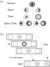
Although attention is distributed across time as well as space, the temporal allocation of attention has been less well researched than its spatial counterpart. A temporal analog of the covert spatial orientation task [Posner MI, Snyder CRR, Davidson BJ (1980) Attention and the detection of signals. J Exp Psychol Gen 109:160-174] was developed to compare the neural systems involved in directing attention to spatial locations versus time intervals. We asked whether there exists a general system for allocating attentional resources, independent of stimulus dimension, or whether functionally specialized brain regions are recruited for directing attention toward spatial versus temporal aspects of the environment. We measured brain activity in seven healthy volunteers by using positron emission tomography (PET) and in eight healthy volunteers by using functional magnetic resonance imaging (fMRI). The task manipulated cued attention to spatial locations (S) and temporal intervals (T) in a factorial design. Symbolic central cues oriented subjects toward S only (left or right), toward T only (300 msec or 1500 msec), toward both S and T simultaneously, or provided no information regarding S or T. Subjects also were scanned during a resting baseline condition. Behavioral data showed benefits and costs for performance during temporal attention similar to those established for spatial attention. Brain-imaging data revealed a partial overlap between neural systems involved in the performance of spatial versus temporal orientation of attention tasks. Additionally, hemispheric asymmetries revealed preferential right and left parietal activation for spatial and temporal attention, respectively. Parietal cortex was activated bilaterally by attending to both dimensions simultaneously. This is the first direct comparison of the neural correlates of attending to spatial versus temporal cues.
Figures


References
-
- Coull JT, Frith CD, Frackowiak RSJ, Grasby PM. A fronto-parietal network for rapid visual information processing: a PET study of sustained attention and working memory. Neuropsychologia. 1996;34:1085–1095. - PubMed
-
- Coull JT, Frackowiak RSJ, Frith CD (1998) Monitoring for target objects: activation of right frontal and parietal cortices with increasing time on task. Neuropsychologia, in press. - PubMed
-
- Deiber M-P, Ibanez V, Sadato N, Hallett M. Cerebral structures participating in motor preparation in humans: a positron emission tomography study. J Neurophysiol. 1996;75:233–247. - PubMed
Publication types
MeSH terms
Grants and funding
LinkOut - more resources
Full Text Sources
