. 1998 Sep 15;18(18):7535-42.
doi: 10.1523/JNEUROSCI.18-18-07535.1998.

Distributed encoding and retrieval of spatial memory in the hippocampus

Affiliations

Distributed encoding and retrieval of spatial memory in the hippocampus

M B Moser et al. J Neurosci. .

Abstract

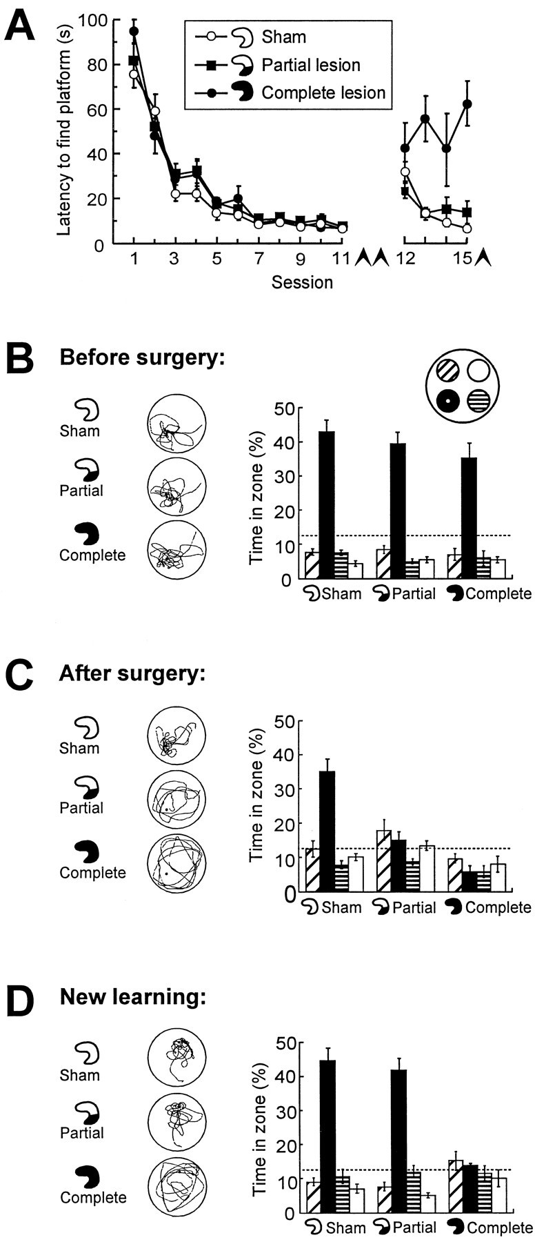
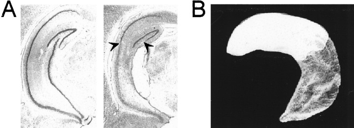
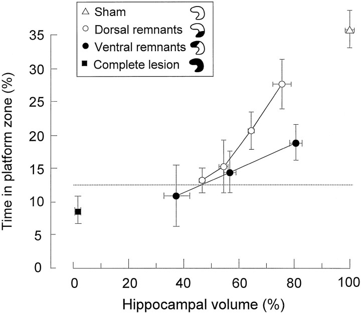
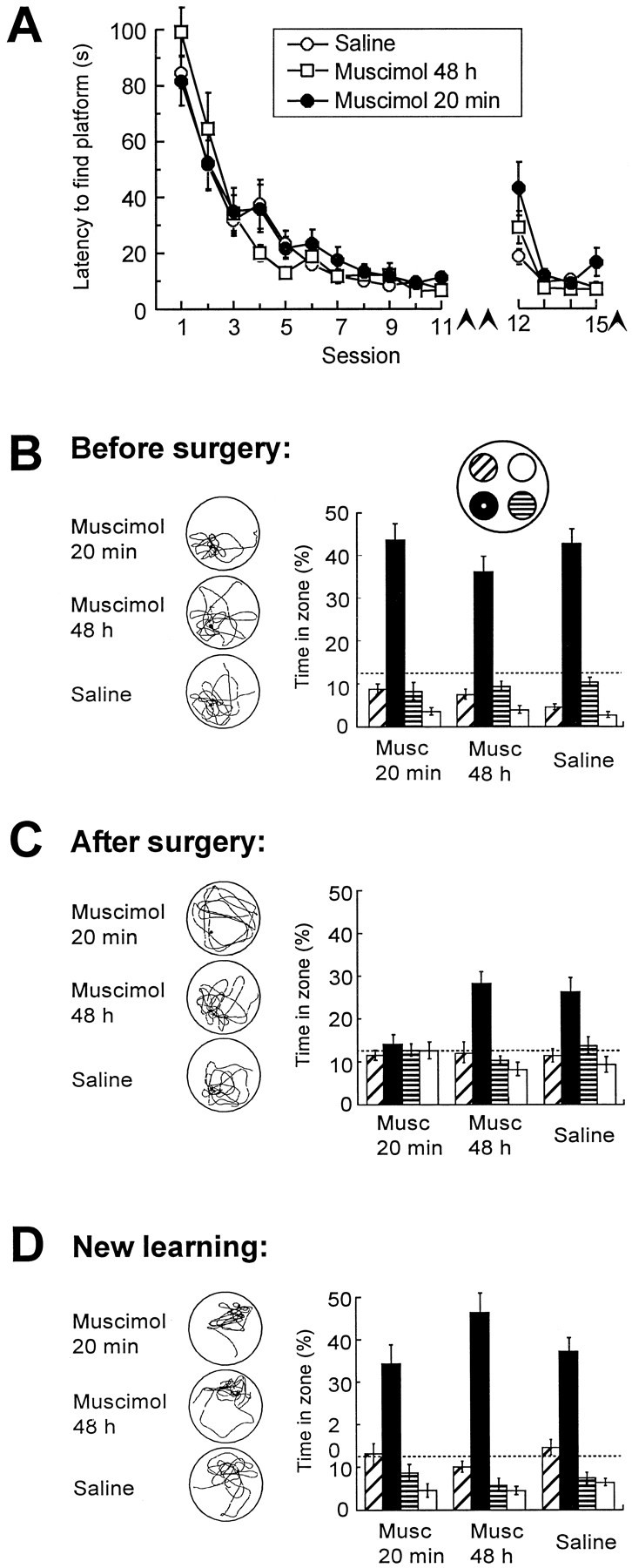
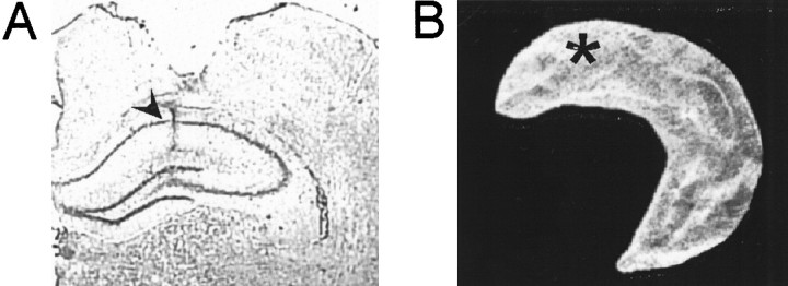
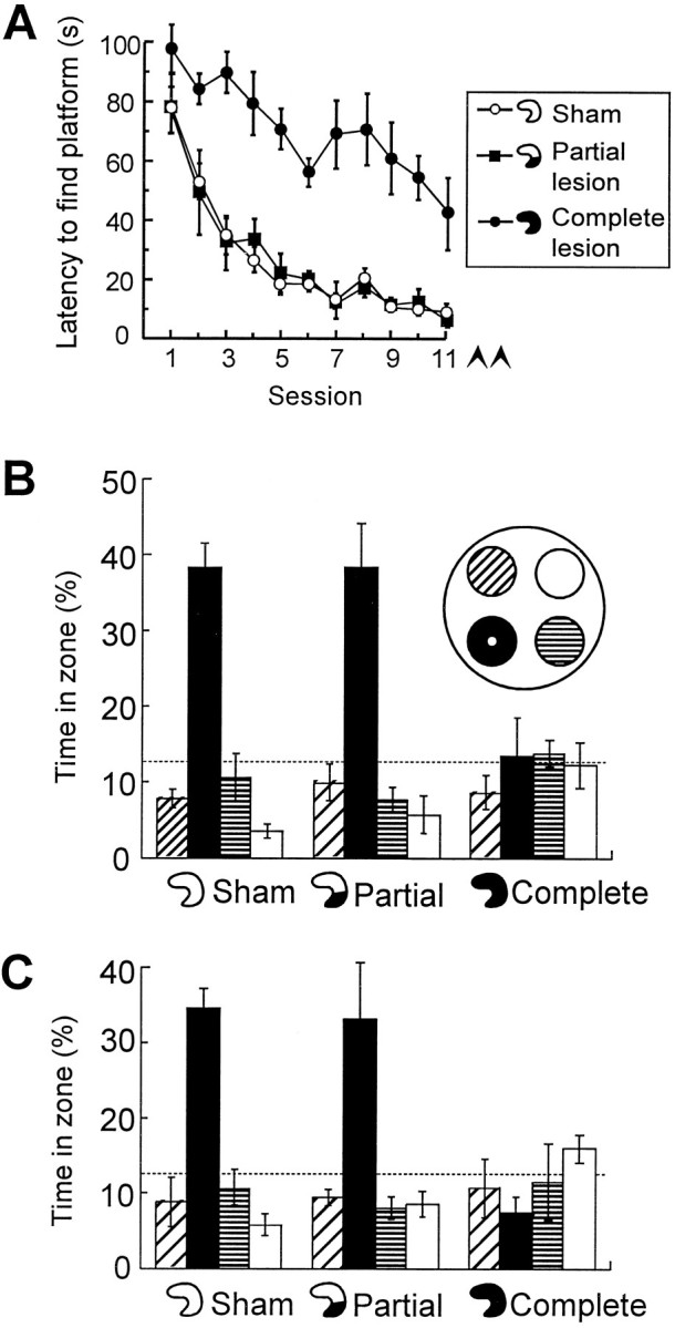
To determine whether memory is processed in a localized or distributed manner by the hippocampus, we inactivated small regions of the structure in pretrained rats before a retention test. Ibotenic acid-induced lesions removing 40% of the hippocampal tissue disrupted retrieval of spatial memory in a water maze but failed to affect new learning or retrieval of a task that was acquired postoperatively. Partial inactivation of the hippocampus by local intrahippocampal 5-aminomethyl-3-hydroxyisoxazole muscimol infusion also impaired retrieval but not new learning. This impairment was temporary; infusions had no effect on retrieval of predrug performance when the test was conducted 48 hr after the infusion. Systematic variation of the volume of dorsal and ventral hippocampal lesions showed that successful retrieval required the integrity of the entire dorsal 70% of the hippocampus. Our data suggest that although spatial tasks can be acquired with local ensembles of hippocampal neurons when other parts of the hippocampus are inactivated, spatial memory is normally both encoded and retrieved by a widely distributed hippocampal network.


Figures

Fig. 1.
Fig. 1.
Retrieval of spatial memory in a water maze after a small hippocampal lesion. Control groups received sham surgery or complete hippocampal lesions. Lesions were induced after 11 sessions of pretraining. Seven days later, retrieval was tested, and the animals were trained in a new task. A, Latency to locate the hidden platform before surgery (sessions 1–11) and after surgery (sessions 12–15, conducted in a new environment).Arrowheads indicate spatial probe tests.B–D, Retrieval on probe tests at the end of pretraining (B), 7 d after pretraining and surgery (C), and after postoperative training in the new environment (D). Left column, Typical swim paths. Right column, Time spent in a circular zone around the platform position (filled bars) and in corresponding zones of the three other quadrants (mean ± SEM). Chance level is 12.5% (dashed lines).
Fig. 2.
Fig. 2.
Location of remaining hippocampal tissue in a rat with a partial hippocampal lesion that impaired retrieval but not new learning (Fig. 1). A, Coronal section showing cresyl violet stains of neuronal cell bodies in the intermediate to ventral portion of the hippocampus of a sham-operated rat (left) and a rat with a lesion that spared 58.5% of hippocampal volume dorsally (right). Arrowheads indicate border between lesioned and healthy tissue. B, Three-dimensional reconstruction of the remaining dorsal hippocampal tissue (white) of the lesioned rat in Asuperimposed on a reconstruction of the entire hippocampus (gray).
Fig. 3.
Fig. 3.
Retrieval on a spatial probe test 7 d after pretraining and surgery as a function of remaining hippocampal volume in animals with dorsal remnants (10% bins) or ventral remnants (20% bins). Retrieval is expressed as the proportion of time spent in a circular zone around the platform position (mean ± SEM).Dashed line indicates chance level.
Fig. 4.
Fig. 4.
Distribution of swim time in pretrained rats after partial inactivation of the dorsal hippocampus by microinfusion of the GABAA receptor agonist muscimol. The interval between pretraining and drug infusion was 7 d. A, Latency to locate the hidden platform during pretraining (sessions 1–11) and after drug infusion (sessions 12–15, conducted in a new environment). Arrowheadsindicate spatial probe tests. B–D, Retrieval on probe tests at the end of preoperative training (B), 7 d after pretraining and surgery (C), and after new learning in a different environment 1.2 hr subsequent to the test in C (D). Muscimol was infused 20 min or 48 hr before the test in C. A separate group received saline 20 min before testing. No drug was given inB. Left column, Typical swim paths.Right column, Time spent in the central zones of each quadrant (Fig. 1). Dashed lines indicate chance level.
Fig. 5.
Fig. 5.
Location of the muscimol infusion within the dorsal hippocampus. A, Cresyl violet stain showing position of internal cannula (arrowhead) in the hippocampus of a representative muscimol-infused rat. The neocortical damage was attributable to the implanted guide cannula. B, Three-dimensional reconstruction showing the position of the cannula (asterisk) in one hippocampus of the same rat.
Fig. 6.
Fig. 6.
Retrieval in animals that received training after the partial hippocampal lesion. The rats had lesions sparing the dorsal hippocampus (as in Fig. 2), complete hippocampal lesions, or sham lesions. A, Latency to locate the hidden platform during postoperative training. Arrowheads indicate spatial probe tests. B, C, Retrieval at the end of training (B) and 7 d later (C). Symbols are the same as in Figure 1.

References

    1. Amaral DG, Witter MP. The three-dimensional organization of the hippocampal formation: a review of anatomical data. Neuroscience. 1989;31:571–591. - PubMed
    1. Andersen P, Bliss TVP, Skrede KK. Lamellar organization of hippocampal excitatory pathways. Exp Brain Res. 1971;13:222–238. - PubMed
    1. Andreasen NC, O’Leary DS, Arndt S, Cizadlo T, Rezai K, Watkins GL, Ponto LLB, Hichwa RD. I. PET studies of memory: novel and practiced free recall of complex narratives. NeuroImage. 1995a;2:284–295. - PubMed
    1. Andreasen NC, O’Leary DS, Cizadlo T, Arndt S, Rezai K, Watkins GL, Ponto LLB, Hichwa RD. II. PET studies of memory: novel versus practiced free recall of word lists. NeuroImage. 1995b;2:296–305. - PubMed
    1. Buckner RL, Goodman J, Burock M, Rotte M, Koutstaal W, Schacter D, Rosen B, Dale AM. Functional-anatomic correlates of object priming in humans revealed by rapid presentation event-related fMRI. Neuron. 1998;20:285–296. - PubMed

Publication types