. 1998 Sep 15;95(19):11065-70.
doi: 10.1073/pnas.95.19.11065.

The 2.8-A structure of rat liver F1-ATPase: configuration of a critical intermediate in ATP synthesis/hydrolysis

Affiliations

The 2.8-A structure of rat liver F1-ATPase: configuration of a critical intermediate in ATP synthesis/hydrolysis

M A Bianchet et al. Proc Natl Acad Sci U S A. .

Abstract

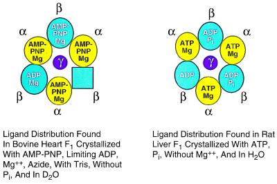
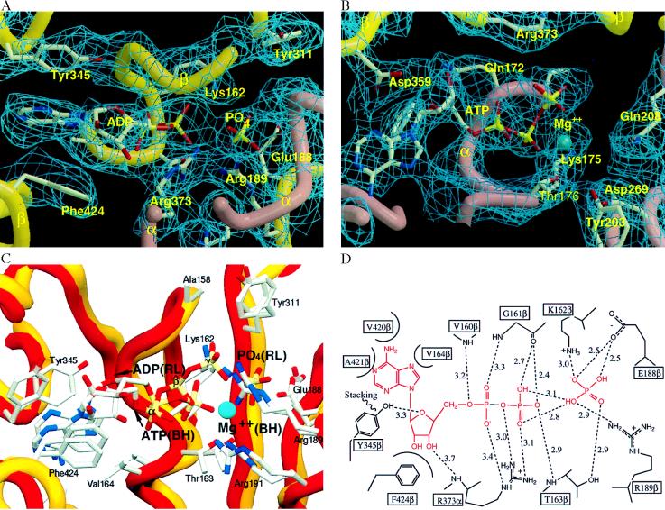
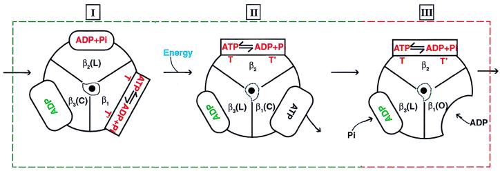
During mitochondrial ATP synthesis, F1-ATPase-the portion of the ATP synthase that contains the catalytic and regulatory nucleotide binding sites-undergoes a series of concerted conformational changes that couple proton translocation to the synthesis of the high levels of ATP required for cellular function. In the structure of the rat liver F1-ATPase, determined to 2.8-A resolution in the presence of physiological concentrations of nucleotides, all three beta subunits contain bound nucleotide and adopt similar conformations. This structure provides the missing configuration of F1 necessary to define all intermediates in the reaction pathway. Incorporation of this structure suggests a mechanism of ATP synthesis/hydrolysis in which configurations of the enzyme with three bound nucleotides play an essential role.


Figures

Figure 1
Figure 1
Schematic representation of the crystallization conditions and the bound nucleotides in the two-nucleotide structure (Left, bovine heart, ref. 14) and the “three-nucleotide structure” (Right, rat liver, this work).
Figure 2
Figure 2
(A) Diffraction quality crystals of rat liver F1. (B) Scan of an SDS/PAGE gel of redissolved F1 crystals. Crystals of F1 were dissolved in 50 μl of 250 mM KPi plus 5 mM EDTA (pH 7.5), and 15 μg were subjected to SDS/PAGE in cylindrical gels. (C) Capacity of redissolved crystals to rebind to F1-depleted inner membrane vesicles of mitochondria and regain sensitivity to oligomycin, an inhibitor of oxidative phosphorylation. Where indicated, 1 μg oligomycin was present. (D) Relative capacity of ATP and MgCl2 to initiate the F1 ATPase reaction. The decrease in optical density of NADH at 340 nm was monitored. Reaction 1 was initiated with 4 mM ATP, and reaction 2 was initiated with 4.8 mM MgCl2.
Figure 3
Figure 3
Ribbon diagram of the rat liver F1-ATPase. Rat liver F1 can be described as an “inverted apple” 110 Å in diameter and 125 Å in length (including the protruding γ subunit) formed by three α and three β subunits alternating around the 3-fold axis of symmetry of the R32 cell, in agreement with the 3.6-Å resolution structure reported (17). The molecule is most narrow at the “top,” where a depression surrounding the 3-fold axis marks the beginning of a large central channel partially occupied by the γ subunit. Parts of the γ subunit extend well beyond the end of the major subunits, forming a long narrow protrusion (the “stem” of the apple). This side, the “bottom” of the F1, is thought to be closest to the membrane in the F0F1 complex. (a) “Side view” of complete model (viewed from a direction perpendicular to the 3-fold axis).The α and the β subunits are also alike, both composed of three domains: an NH2-terminal β sheet domain, a nucleotide binding domain, and a COOH-terminal domain. (b) “Top view” of the NH2-terminal domain. (c) Top view of the nucleotide binding domain. (d) Top view of the COOH-terminal domain. In the top views (c and d) the corresponding portions of the γ subunit are shown in yellow.
Figure 4
Figure 4
Differences in position of the γ subunit between the rat liver and the bovine heart enzyme. The lower part of the γ subunit in rat liver F1 (gold) is displaced from the central axis ≈4 Å more than the bovine heart γ subunit (red); bovine heart subunits βTPE (opposite subunit pair) were used as references for alignment.
Figure 5
Figure 5
Nucleotide binding sites of rat liver F1-ATPase. (A) Electron density of the β subunit binding site. The main chain of β is shown in gold, and that of α is shown in rose. Oxygens are red; all other atoms in gray. (B) Electron density of α subunit binding site. Color scheme is as in A, and Mg2+ is in blue. The adenine ring binds in a pocket making van der Waals contacts as well as hydrogen bonding interactions with the protein. Main chain and side atoms provide interactions to the ribose, the phosphates, and the Mg2+. (C) Comparison of the β subunit nucleotide binding site of rat liver F1 (gold) to that of the βT subunit of bovine heart F1 (red). (D) Schematic diagram of nucleotide binding to the β subunit. Close interactions and hydrogen bonds (with distances in Å) are shown. The ADP and the Pi are shown in red.
Figure 6
Figure 6
Proposed mechanism for ATP synthesis/hydrolysis. In the figure, β subunits in one F1 molecule are labeled β1, β2, and β3. Two configurations of the enzyme are sufficient to describe all of the intermediates in the proposed reaction pathway: (i) the three-nucleotide structure with T (tight), L (loose), and C (closed) sites in the β subunits (I and II, boxed by the green dashed line); and (ii) the two-nucleotide structure with T, L, and O (open) sites (III, boxed by the red dashed line). Conformations T and T′ of the β subunit are highly similar, but T contains ATP and T′ contains ADP plus Pi. Starting with the most stable species (I), ATP synthesis proceeds through a proton translocation driven conformational change. This transformation (I to II) can occur only with ATP in the T site. Next, ATP is released from the C site which, after release, changes to the O conformation. After intermediate III is reached, the molecule binds ADP in the O site, which immediately changes to the C-conformation, and Pi in the L site becomes species I (but with subunit numbers cyclically permuted). The reaction proceeds cyclically through these steps. Note that, in this mechanism, in contrast to previous proposals (4, 6, 9, 14), the concerted conformational change involving all three β subunits (I to II) occurs in the three nucleotide form of the enzyme and does not involve a β subunit in the open conformation.

References

    1. Catterall W A, Pedersen P L. J Biol Chem. 1971;246:4987–4994. - PubMed
    1. Catterall W A, Coty W A, Pedersen P L. J Biol Chem. 1973;248:7427–7431. - PubMed
    1. Catterall W A, Pedersen P L. Biochem Soc Spec Publ. 1974;4:63–88.
    1. Boyer P D. Annu Rev Biochem. 1997;66:714–749. - PubMed
    1. Weber J, Senior A E. Biochim Biophys Acta. 1997;1319:19–58. - PubMed

Publication types

MeSH terms

Substances