. 1998 Sep 15;95(19):11146-51.
doi: 10.1073/pnas.95.19.11146.

Unexpected frameshifts from gene to expressed protein in a phage-displayed peptide library

Affiliations

Unexpected frameshifts from gene to expressed protein in a phage-displayed peptide library

J Cárcamo et al. Proc Natl Acad Sci U S A. .

Abstract

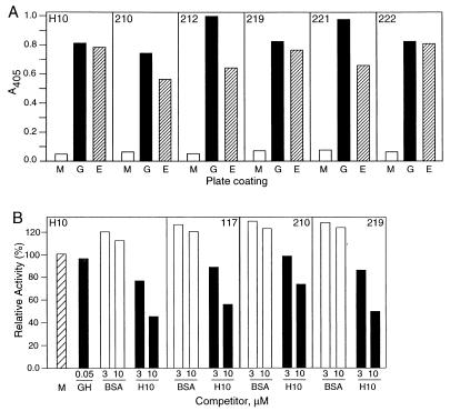
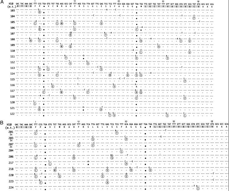
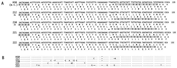
A library of long peptides displayed on the pIII protein of filamentous phage was used in biopanning experiments against several protein targets. We find that a large percentage of phage clones that bind specifically to a target contain peptide-encoding genes that do not have an ORF. Instead, the reading frame is either interrupted by one or more nonsuppressed stop codons, or a post-transcriptional frameshift is needed to account for the expression of the minor phage coat protein pIII. The percentage of frameshifted clones varies depending on the target. It can be as high as 90% for clones specific for soluble forms of certain cytokine receptors. Conversely, biopanning against four mAbs did not yield any frameshifted clones. Our studies focused on one clone that binds specifically to rat growth hormone binding protein (GHBP) yet does not have an ORF. A secondary peptide library containing random mutations of this sequence was constructed and panned against GHBP to optimize and correct the reading frame. In the last round (round two) of panning with this library, none of the phage clones that bound to GHBP had an ORF. However, careful analysis of these clones allowed us to design a synthetic peptide capable of binding to GHBP. The results of this study indicate that ORFs are not required to obtain gene expression of the minor coat protein of filamentous phage and suggest that some ORF- clones may have a selective advantage over the clones having ORFs.


Figures

Figure 1
Figure 1
ELISA analysis of phage binders to GHBP. (A) Binding of the H10 phage or its round-two frameshifting mutants to GHBP, anti-E-tag mAb, or nonfat milk-coated plates. Wells were coated with either 2% nonfat milk (M), 100 ng/well of GHBP (G), or 100 ng/well of anti-E-tag mAb (E). Phage were added at 1010/well and were detected by anti-M13 antibody–horseradish peroxidase conjugate. The clone name is indicated in the upper portions of the graphs. (B) Competition of H10 phage binding. Phage were added at 1010/well in the absence or presence of competitor. Competitors were GH, BSA, or the H10 synthetic peptide, added 1 h before phage. Phage were detected by anti-M13 antibody–horseradish peroxidase conjugate. Relative activity is defined as the ratio of ELISA A405 values in the presence and the absence of the competitor. The A405 for the control (no competitor) was 0.4 and is labeled M in the graph.
Figure 2
Figure 2
Sequences of H10 mutants. (A) Results of panning round one. (B) Frame 0 sequences from round two. Deduced N-terminal FLAG sequence and the synthetic peptide binder sequence are shaded. Large black dots highlight nonsuppressed stop codons. Sequence differences from the wild-type H10 sequence are shown, and sequence identities are indicated by dashes. Amino acid replacements are shown in circles. The clone number is listed on the left side of the figure. Symbol “2x” under the clone number indicates the number of occurrences of this clone. The UAG codons are translated as Gln in the table because the termination of the translation is suppressed in the E. coli strain used.
Figure 3
Figure 3
Sequences of frameshifted H10 mutants (round two). A shows DNA sequences and three reading frames. The FLAG and deduced peptide binder sequences are shaded. Other symbols are as in Fig. 2. B shows a DNA sequence alignment. The alignment compares the H10 sequence with the 221, 222, 212, and 210 sequences. Dots indicate identities, and dashes indicate deletions. The UAG codons are translated as Gln in the figure because the termination of the translation is suppressed in the E. coli strain used.

Similar articles

Cited by

References

    1. Scott J K, Smith G P. Science. 1990;249:386–390. - PubMed
    1. Grihalde N D, Chen Y C, Golden A, Gubbins E, Mandecki W. Gene. 1995;166:187–195. - PubMed
    1. Chen Y C J, Delbrook K, Dealwis C, Mimms L, Mushawar I K, Mandecki W. Proc Natl Acad Sci USA. 1996;93:1997–2001. - PMC - PubMed
    1. Kay B K, Adey N B, He Y S, Manfredi J P, Mataragnon A H, Fowlkes D M. Gene. 1993;128:59–65. - PubMed
    1. Lowman H B. Annu Rev Biophys Biomol Struct. 1997;26:401–424. - PubMed

MeSH terms

LinkOut - more resources