. 1998 Sep 15;95(19):11181-6.
doi: 10.1073/pnas.95.19.11181.

An essential role for the phosphatidylinositol transfer protein in the scission of coatomer-coated vesicles from the trans-Golgi network

Affiliations

An essential role for the phosphatidylinositol transfer protein in the scission of coatomer-coated vesicles from the trans-Golgi network

J P Simon et al. Proc Natl Acad Sci U S A. .

Abstract

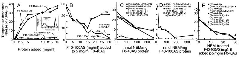
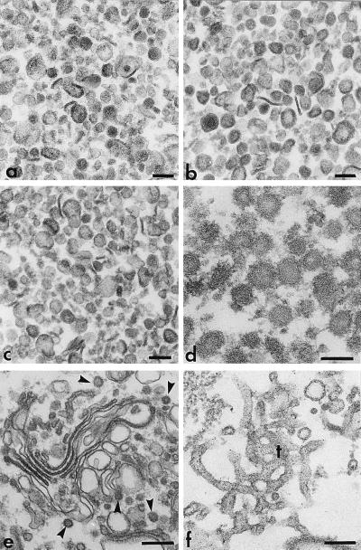
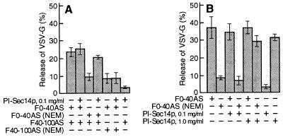
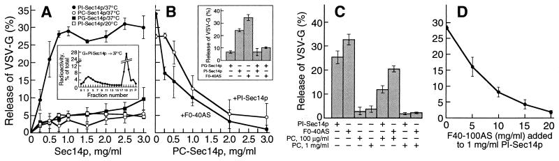
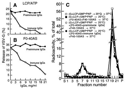
We identified the phosphatidylinositol transfer protein (PITP) as being responsible for a powerful latent, nucleotide-independent, Golgi-vesiculating activity that is present in the cytosol but is only manifested as an uncontrolled activity in a cytosolic protein subfraction, in which it is separated from regulatory components that appear to normally limit its action to the scission of COPI-coated buds from trans-Golgi network membranes. A specific anti-PITP antibody that recognizes the two mammalian PITP isoforms fully inhibited the capacity of the cytosol to support normal vesicle generation as well as the uncontrolled vesiculating activity manifested by the cytosolic protein subfraction. The phosphatidylinositol- (PI) loaded form of the yeast PITP, Sec14p, but not the phosphatidylcholine- (PC) loaded form of the protein, was capable of substituting for the cytosolic subfraction in promoting the scission of coated buds from the trans-Golgi network. At higher concentration, however, Sec14p, when loaded with PI, but not with PC or phosphatidylglycerol, caused by itself an indiscriminate vesiculation of uncoated Golgi membranes that could be suppressed by PC-Sec14p, which also suppresses the uncontrolled vesiculation caused by the cytosolic subfraction. We propose that, by delivering PI to specific sites in the Golgi membrane near the necks of coated buds, PITP induces local changes in the organization of the lipid bilayer, possibly involving PI metabolites, that triggers the fusion of the ectoplasmic faces of the Golgi membrane necessary for the scission of COPI-coated vesicles.


Figures

Figure 1
Figure 1
A latent, NEM-sensitive, Golgi-vesiculating activity that participates in normal vesicle generation is present in the cytosol. (A) Nucleotide-independent Golgi-vesiculating activity of the F0–40AS subfraction. Golgi fractions were incubated for 60 min at either 37°C or 4°C, with (+EN) or without (−EN) an energy-generating system, in the presence of varying concentrations of either the F0–40AS or F40–100AS cytosolic subfractions and the temperature-dependent release of labeled VSV-G protein was measured. Inset, Sucrose gradient profile of labeled VSV-G protein released during an incubation with F0–40AS (15 mg/ml). Each point represents the average from four experiments using different preparations of F0–40AS. (B) The F40–100AS subfraction suppresses the nucleotide-independent vesiculating activity of F0–40AS and, when recombined with it, restores nucleotide-dependent vesicle generation (+EN). Assays were carried out as in A with F0–40AS (5 mg/ml) supplemented with varying amounts of the F40–100AS, with or without an energy-generating system, as indicated. (C and D) The nucleotide-dependent formation of TGN-derived vesicles requires at least two NEM-sensitive cytosolic factors, one of which is also necessary for the nucleotide-independent process of vesicle release. (C) Aliquots of F0–40AS were treated (10 min at 0°C) with varying amounts of NEM followed by addition of a 2-fold molar excess of DTT. They were then used (5 mg/ml) to support vesicle release with or without F40–100AS (25 mg/ml) in the presence or absence of an energy supply. (D) F40–100AS was treated with varying amounts of NEM, as described in C, and used (25 mg/ml) to support vesicle release, with or without the addition of F0–40AS (5 mg/ml) in the presence or absence of an ATP supply. (E) The suppressing factor in F40–100AS is not sensitive to NEM. F40–100AS was treated (0°C for 10 min) with NEM (175 nmol NEM/mg protein) or mock treated with control buffer, followed by the addition of a 2-fold molar excess of DTT. Vesicle release was measured after incubation of the Golgi membranes with various concentrations of NEM-treated or control F40–100AS, with or without F0–40AS (5 mg/ml), in the presence or absence of an ATP supply as indicated.
Figure 2
Figure 2
Naked and coated vesicles released in vitro by PITP. (a–c) Naked vesicles generated from Golgi membranes by incubation (37°C, 60 min) with total liver cytosolic proteins and ATP (a), F0–40AS (5 mg/ml, b), or PI-Sec14p (1 mg/ml, c). The vesicles were purified as previously described (28). (d) Purified COPI-coated vesicles released from Golgi membranes that, after priming for coat assembly/bud formation (14), were reincubated at 37°C for vesicle scission with PI-Sec14p (1 mg/ml) and F40–100AS (25 mg/ml). (e and f) Whereas incubation (30 min at 20°C) of Golgi membranes for coat assembly with liver cytosolic proteins and guanylyl-imidodiphosphate (100 μM) leads to the production of coated buds (arrowheads) on stacked cisternae (e), a similar incubation with PI-Sec14p (1 mg/ml) leads to extensive tubulation of the Golgi membranes (f). All samples were recovered by sedimentation (10,000 × g for 10 min), fixed with 1% glutaraldehyde and OsO4, stained with tannic acid, and processed for routine electron microscopy. Bars, 100 nm (a–d) or 200 nm (e and f).
Figure 3
Figure 3
The NEM-sensitive factor in F0–40AS can be replaced by the yeast PITP, Sec14p. (A) Cytosolic protein subfractions were mock-treated or treated with NEM (350 nmol NEM/mg protein for F0–40AS and 75 nmol NEM/mg protein for F40–100AS) and used as indicated (2 mg/ml of F0–40AS and/or 8 mg/ml of F40–100AS), with or without PI-Sec14p (0.1 mg/ml), for ATP-dependent vesicle generation. (B) Nucleotide-independent vesicle generation assays were carried out in the presence or absence of the indicated concentrations of PI-Sec14p with either mock-treated (F0–40AS) or NEM-treated (350 nmol NEM/mg protein) F0–40AS (5 mg/ml). In A and B, each point represents the mean (±SD) of four determinations.
Figure 4
Figure 4
PI-Sec14p vesiculates TGN membranes. (A) Golgi membranes (0.25 mg/ml) were incubated (60 min at 37°C or 20°C, as indicated) with varying concentrations of Sec14p charged with either PI, PC, or PG. Inset, Sucrose gradient profile of released VSV-G protein after incubation with PI-Sec14p (1 mg/ml). (B) PC- and PG-charged Sec14p prevent the vesiculation caused by PI-Sec14p (1 mg/ml) or F0–40AS (5 mg/ml). Vesicle generation reactions were carried out in the absence of nucleotides with either PI-Sec14p or F0–40AS and the indicated concentrations of PC-Sec14p. Inset, Incubations were carried out as in A with either PI-Sec14p (1 mg/ml) or F0–40AS (5 mg/ml) in the presence or absence of PG-Sec14p (3 mg/ml). (C) The addition of PC-containing liposomes to PI-Sec14p (1 mg/ml) or to F0–40AS (5 mg/ml) abolishes their capacity to vesiculate Golgi membranes, as expected from the replacement of PI in PITP by PC. Vesicle release assays were carried out with PI-Sec14p (1 mg/ml) or F0–40AS fraction (5 mg/ml) and the indicated concentrations of PC-containing liposomes. (D) F40–100AS abolishes the PI-Sec14p-mediated vesiculation of TGN membranes. Incubations for vesicle generation were carried out with PI-Sec14p (1 mg/ml) and various amounts of F40–100AS. (A–D) Each point represents the average (±SD) from three determinations using different Sec14p preparations.
Figure 5
Figure 5
PITP functions in vesicle scission. (A) Scission of coated buds requires at least two cytosolic components, one in F0–40AS and the other in F40–100AS. In a two-step vesicle generation assay, Golgi membranes (G) were incubated (30 min at 20°C) for coat assembly/bud formation with total liver cytosolic protein (LCP; 10 mg/ml) and guanylyl-imidodiphosphate (100 μM). The primed Golgi membranes were recovered and reincubated (60 min at 37°C) with either F0–40AS (2 mg/ml, open circles) or F40–100AS (8 mg/ml, closed circles) or a combination of both (open squares). The samples were then analyzed by sucrose gradient centrifugation. The values are the averages from six independent experiments. (B) Scission of coated buds requires an NEM-sensitive activity present in F0–40AS. Golgi membranes, primed as in A, were reincubated (60 min at 37°C) with a combination of mock- or NEM-treated (350 nmol NEM/mg protein) F0–40AS (2 mg/ml) and mock- or NEM-treated (175 nmol NEM/mg protein) F40–100AS (8 mg/ml), as indicated. Vesicle release was determined as in A, in four independent experiments. (C) F0–40AS can be replaced in the scission reaction by PI-Sec14p. Primed Golgi membranes were reincubated as in A, but with F40–100AS (20 mg/ml) either alone (closed squares) or along with either PI-Sec14p (1 mg/ml, open circles), PC-Sec14p (1 mg/ml, closed circles), or PI-containing liposomes (1 mg/ml, open squares). Each point represents the average from three (closed circles) or nine (other symbols) independent experiments using three different preparations of Sec14p. (Inset) Extent of coated vesicle scission as a function of PI-Sec14p concentration. (D) Vesiculation of uncoated portions of primed Golgi membranes by PI-Sec14p. Primed Golgi membrane fractions were incubated with either PI-Sec14p (1 mg/ml, open triangles), PC-Sec14p (1 mg/ml, closed triangles), or buffer alone (open circles). Points represent averages from three (closed triangles) or nine (other symbols) independent experiments using three different Sec14p preparations.
Figure 6
Figure 6
(A and B) Anti-PITPα antibodies inhibit the production of VSV-G-containing Golgi vesicles. (A) Assays were carried out with total liver cytosolic proteins (LCP; 10 mg/ml) and 1 mM ATP or with F0–40AS (5 mg/ml, B) pretreated for 60 min at 4°C with various concentrations of anti-PITPα or preimmune IgG from sera of a rabbit immunized with recombinant PITPα (35). Vesicle release is plotted as a function of the final concentration of IgG. (C) Anti-PITPα antibodies prevent the scission of COPI-coated vesicles from the TGN. Primed Golgi membranes were reincubated (60 min at 37°C) for coated vesicle scission with either buffer alone (open squares), a combination of F40–100AS (8 mg/ml) and F0–40AS (2 mg/ml, filled squares), or a combination of the two subfractions in which the F0–40AS had been preincubated (60 min at 4°C) with preimmune (open circles) or immune (filled circles) IgGs (20 mg/ml). The reaction mixtures were analyzed as in Fig. 5.

Similar articles

Cited by

References

    1. Barlowe C, Orci L, Yeung T, Hosobuchi M, Hamamoto S, Salama N, Rexach M F, Ravazzola M, Amherdt M, Schekman R. Cell. 1994;77:895–907. - PubMed
    1. Serafini T, Stenbeck G, Brecht A, Lottspeich F, Orci L, Rothman J E, Wieland F T. Nature (London) 1991;349:215–220. - PubMed
    1. Donaldson J G, Finazzi D, Klausner R D. Nature (London) 1992;360:350–352. - PubMed
    1. Traub L M, Osatrom J A, Kornfeld S. J Cell Biol. 1993;123:561–573. - PMC - PubMed
    1. Helms J B, Rothman J E. Nature (London) 1992;360:352–354. - PubMed

Publication types

MeSH terms