Case Reports
. 1998 Sep 15;95(19):11312-7.
doi: 10.1073/pnas.95.19.11312.

A dysfunctional desmin mutation in a patient with severe generalized myopathy

Affiliations
Case Reports

A dysfunctional desmin mutation in a patient with severe generalized myopathy

A M Muñoz-Mármol et al. Proc Natl Acad Sci U S A. .

Abstract

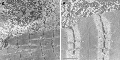
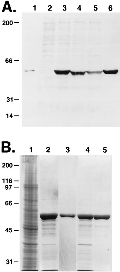
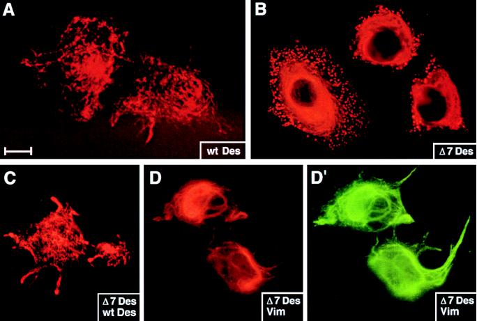
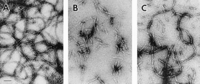
Mice lacking desmin produce muscle fibers with Z disks and normal sarcomeric organization. However, the muscles are mechanically fragile and degenerate upon repeated contractions. We report here a human patient with severe generalized myopathy and aberrant intrasarcoplasmic accumulation of desmin intermediate filaments. Muscle tissue from this patient lacks the wild-type desmin allele and has a desmin gene mutation encoding a 7-aa deletion within the coiled-coil segment of the protein. We show that recombinant desmin harboring this deletion cannot form proper desmin intermediate filament networks in cultured cells, nor is it able to assemble into 10-nm filaments in vitro. These findings provide direct evidence that a mutation in desmin can cause human myopathies.


Figures

Figure 1
Figure 1
Ultrastructural abnormalities in a skeletal muscle biopsy. Note splitting of myofibrils and aberrations in Z band register. Subsarcolemmal aggregates of granular and filamentous material were prevalent. The aggregates show the same electron density as the Z bands, and are in continuity with them. Similar ultrastructural images were seen in myocardium and in small intestine muscularis propria (18). (Bar represents 1 μm.)
Figure 2
Figure 2
(A) Pedigree of the desmin-related myopathy family. Family members from whom DNA samples were obtained are numbered. Squares: males. Circles: females. Solid symbol: patient. Open symbols: wild-type individuals. Semiopen symbols: carriers of the deletion. Crossed symbol: deceased individuals. (B) SSCP analysis showing the different single-strand band pattern of exon 1 (4FR) amplimers from the proband (II.1) and a wild-type control individual (WT). (C) Exon 1 amplimers from the available family members and a control individual (WT) run in a denaturing acrylamide gel to detect wild-type (437 nt) and mutant (416 nt) alleles.
Figure 3
Figure 3
Location of the putative deletion in relation to desmin’s secondary structure. IF proteins share a common secondary structure consisting of four α-helical, coiled-coil rod segments 1A, 1B, 2A, and 2B, separated by short nonhelical linker sequences (thin lines) and flanked by nonhelical head and tail segments (29). The human desmin amino acid sequence corresponding to the end of helix 1A (amino acids 133–143), linker L1 (amino acids 144–151), and the beginning of helix 1B (amino acids 152–181) are shown. The A135 (see text) and the seven deleted residues (173–179) are highlighted (28). Arrow indicates approximate localization of the deletion.
Figure 4
Figure 4
(A) Antidesmin immunoblot analysis of protein extracts. Proteins were from mouse heart muscle (lane 1), MCF-7 cells transfected with: pCMV (lane 2), pCMVDes (lanes 3 and 5), pCMVΔ7Des (lane 4), pJVim (lane 5), or COS cells transfected with pCMVDes (lane 6). (B) SDS/PAGE analyses of bacterially expressed desmin proteins. Escherichia coli strain BL21(DE3)pLysS transformed with pETDes or pETΔ7Des were harvested and inclusion body fractions (IBFs) were isolated and used for purification of recombinant desmin proteins by Mono-Q anion exchange chromatography. Protein samples were resolved by SDS/PAGE and visualized by Coomassie blue stain. Samples are from: lane 1, total bacterial extract (from pETDes transformed cells); lanes 2 and 4, IBF insoluble protein extracts from pETDes or pETΔ7Des-transformed cells; lanes 3 and 5, purified Des or Δ7Des after Mono-Q anion exchange chromatography. Molecular mass standards are at left in kDa.
Figure 5
Figure 5
De novo desmin IF network formation in MCF-7 cells. Cells were transfected with pCVM (not shown), pCMVDes (A and C), pCMVΔ7Des (B–D′), or pJVim (D and D′). After transient expression of the transgenes, cells were fixed and stained with antibodies against desmin (A–C and D′) or vimentin (D). Shown are representative examples of transfected cells. Under these conditions, no staining was observed in pCVM-transfected or in untransfected cells. (Bar represents 15 μm in A and B, 20 μm in CD′.)
Figure 6
Figure 6
In vitro assembly of wild-type and mutant desmin. Proteins were purified and subjected to in vitro filament assembly (see Materials and Methods). (A) Desmin. (B) Δ7 Desmin. (C) 1:1 mixture of desmin +Δ7 desmin. (Bar represents 100 nm.)

Similar articles

Cited by

References

    1. Li Z, Marchand P, Humbert J, Babinet C, Paulin D. Development (Cambridge, UK) 1993;117:947–959. - PubMed
    1. Gard D L, Lazarides E. Cell. 1980;19:263–275. - PubMed
    1. Lazarides E, Granger B L, Gard D L, O’Connor C M, Breckler J, Price M, Danto S I. Cold Spring Harbor Symp Quant Biol. 1982;46:351–378. - PubMed
    1. Lazarides E, Hubbard B D. Proc Natl Acad Sci USA. 1976;73:4344–4348. - PMC - PubMed
    1. Fuchs E, Cleveland D. Science. 1998;279:514–519. - PubMed

Publication types