. 1998 Sep 15;95(19):11324-9.
doi: 10.1073/pnas.95.19.11324.

Human CUL-1 associates with the SKP1/SKP2 complex and regulates p21(CIP1/WAF1) and cyclin D proteins

Affiliations

Human CUL-1 associates with the SKP1/SKP2 complex and regulates p21(CIP1/WAF1) and cyclin D proteins

Z K Yu et al. Proc Natl Acad Sci U S A. .

Abstract

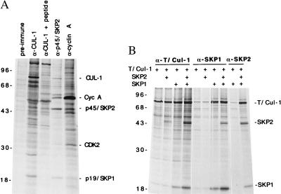
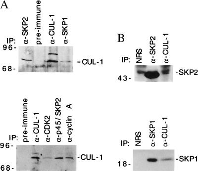
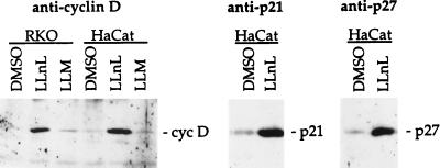
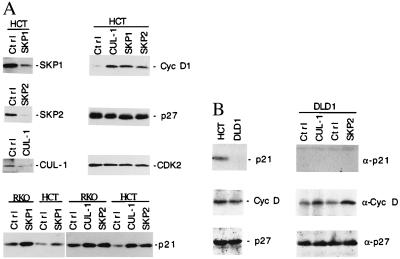
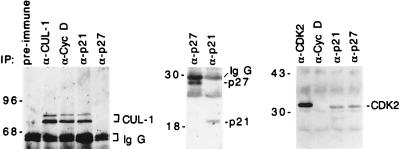
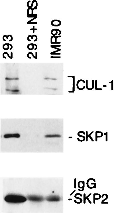
Deregulation of cell proliferation is a hallmark of cancer. In many transformed cells, the cyclin A/CDK2 complex that contains S-phase kinase associated proteins 1 and 2 (SKP1 and SKP2) is highly induced. To determine the roles of this complex in the cell cycle regulation and transformation, we have examined the composition of this complex. We report here that this complex contained an additional protein, human CUL-1, a member of the cullin/CDC53 family. The identification of CUL-1 as a member of the complex raises the possibility that the p19(SKP1)/p45(SKP2)/CUL-1 complex may function as the yeast SKP1-CDC53-F-box (SCF) protein complex that acts as a ubiquitin E3 ligase to regulate the G1/S transition. In mammalian cells, cyclin D, p21(CIP1/WAF1), and p27(KIP1) are short-lived proteins that are controlled by ubiquitin-dependent proteolysis. To determine the potential in vivo targets of the p19(SKP1)/p45(SKP2)/CUL-1 complex, we have used the specific antisense oligodeoxynucleotides against either SKP1, SKP2, or CUL-1 RNA to inhibit their expression. Treatment of cells with these oligonucleotides caused the selective accumulation of p21 and cyclin D proteins. The protein level of p27 was not affected. These data suggest that the human p19(SKP1)/p45(SKP2)/CUL-1 complex is likely to function as an E3 ligase to selectively target cyclin D and p21 for the ubiquitin-dependent protein degradation. Aberrant expression of human p19(SKP1)/p45(SKP2)/CUL-1 complex thus may contribute to tumorigenesis by regulating the protein levels of G1 cell cycle regulators.


Figures

Figure 1
Figure 1
Association between human CUL-1 and p45SKP2/p19SKP1. (A) 293 cells were metabolically labeled with [35S]methionine. The CUL-1, p45SKP2, and cyclin A complexes were isolated by immunoprecipitation using their specific antibodies from labeled 293 cell lysates. The proteins in each protein complex were resolved in an SDS/polyacrylamide gel and compared. The positions of p45SKP2 associated p19SKP1, cyclin A, CDK2, and CUL-1/p86 are indicated on the right. The molecular mass standard (in kDa) are shown on the left. Preimmune serum is the serum from the CUL-1 rabbit before immunization and is used as a control. Peptide, the CUL-1 antigen was included for competition during immunoprecipitation. (B) Human CUL-1 gene was tagged with T7 epitope tag (T/Cul-1) and cloned under the cytomegalovirus promoter control. This construct was cotransfected with expression constructs containing either p19SKP1, p45SKP2, or both into 293 cells as indicated. Twenty-four hours after transfection, the cells were [35S]-methionine-labeled, and the protein complexes were isolated with either T7-tag, p19SKP1-, or p45SKP2-specific antibodies as indicated.
Figure 2
Figure 2
Association between the endogenous CUL-1 and p19SKP1, p45SKP2, cyclin A, and CDK2. (A) Presence of CUL-1 in the p19SKP1, p45SKP2, cyclin A, and CDK2 complexes. The p19SKP1, p45SKP2, CUL-1, cyclin A, and CDK2 complexes were immunoprecipitated from lysates prepared from actively growing HCT116 (Upper) or 293 (Lower) cells by specific antibodies as indicated on the top of each lane, respectively. The presence of CUL-1 is detected by immunoblotting with a CUL-1 specific antibody. The preimmune serum is the same as described in Fig. 1. (B) Presence of p45SKP2 and p19SKP1 in CUL-1 complexes. The CUL-1, p45SKP2, or p19SKP1 complex was immunoprecipitated from HCT116 cell lysates and immunoblotted with either p45SKP2 (Upper) or p19SKP1 (Lower) specific antibodies. NRS, normal rabbit serum from an unrelated rabbit.
Figure 3
Figure 3
Regulation of cyclin D, p21, and p27 by the ubiquitin-dependent proteolysis. (A) Log-phase RKO or HaCat cells were treated with either dimethyl sulfoxide (DMSO) (0.05%), 10 μg/ml of LLNL, or N-acetyl-l-leucinyl-l-leucinyl-methional (LLM) for 6 hr. The cyclin D, p21, and p27 protein were isolated and detected by their respective antibodies by using immunoprecipitation and Western blot analyses.
Figure 4
Figure 4
Selective stabilization of the cellular cyclin D and p21 proteins by antisense oligodeoxynucleotides against SKP1, SKP2, or CUL-1. (A, Upper Left) HCT116 (HCT) cells were transfected with antisense oligonucleotides against SKP1, SKP2, CUL-1, or a control oligonucleotide (Ctrl) by using cytofectin GS2888 as indicated on top of each lane. Eighteen hours after transfection, each protein was immunoprecipitated and quantitated by immunoblotting analysis using the specific antibody as indicated on the right, respectively. (A, Upper Right and Lower) HCT116 or RKO cells were treated with control or antisense oligonucleotides against CUL-1, SKP2, or SKP1 RNA as indicated on the top of each panel. The relative protein levels of cyclin D, p27, CDK2, and p21 from the treated HCT116 or RKO cells (0.5 × 10⁶ each) were compared by immunoprecipitation and immunoblotting analyses using their respective antibodies. (B, Left) The relative levels of p21, cyclin D, and p27 from HCT116 (p53 positive) and DLD1 (p53 negative) cells (0.5 × 10⁶ each) were compared by using immunoprecipitation and Western blotting analyses. (B, Right) DLD1 cells were transfected with antisense oligonucleotides against SKP1, SKP2, CUL-1, or a control oligonucleotide (Ctrl) as indicated on the top of the lanes. The antisense effects on the protein levels of p21, cyclin D, and p27 were examined by the Western blot analysis as described in A.
Figure 5
Figure 5
Human CUL-1 protein associates with cyclin D and p21. Cell lysates from actively growing HaCat cells were immunoprecipitated with specific antibodies as indicated on the top of each lane. The immunoprecipitated protein complexes were blotted with CUL-1 (Left), p27 and p21 (Middle), or CDK2 (Right) antibodies, respectively, as indicated on the right side of each panel.
Figure 6
Figure 6
p45SKP2 is highly induced in transformed cells. Cell lysates (0.5 mg proteins) were made either from actively growing 293, an adenovirus transformed human embryo cell, or from IMR90, a nontransformed human fibroblast. The relative amounts of CUL-1, p19SKP1, and p45SKP2 were determined by immunoprecipitation and Western blot analyses.

Similar articles

Cited by

References

    1. Hunter T, Pines J. Cell. 1994;79:573–582. - PubMed
    1. Sherr C J. Science. 1996;274:1672–1677. - PubMed
    1. Zhang H, Xiong Y, Beach D. Mol Biol Cell. 1993;4:897–906. - PMC - PubMed
    1. Zhang H, Kobayashi R, Galaktionov K, Beach D. Cell. 1995;82:915–925. - PubMed
    1. Bai C, Sen P, Hofmann K, Ma L, Goebl M, Harper J W, Elledge S J. Cell. 1996;86:263–274. - PubMed

Publication types

LinkOut - more resources