. 1998 Sep 15;95(19):11336-41.
doi: 10.1073/pnas.95.19.11336.

Attractin (DPPT-L), a member of the CUB family of cell adhesion and guidance proteins, is secreted by activated human T lymphocytes and modulates immune cell interactions

Affiliations

Attractin (DPPT-L), a member of the CUB family of cell adhesion and guidance proteins, is secreted by activated human T lymphocytes and modulates immune cell interactions

J S Duke-Cohan et al. Proc Natl Acad Sci U S A. .

Abstract

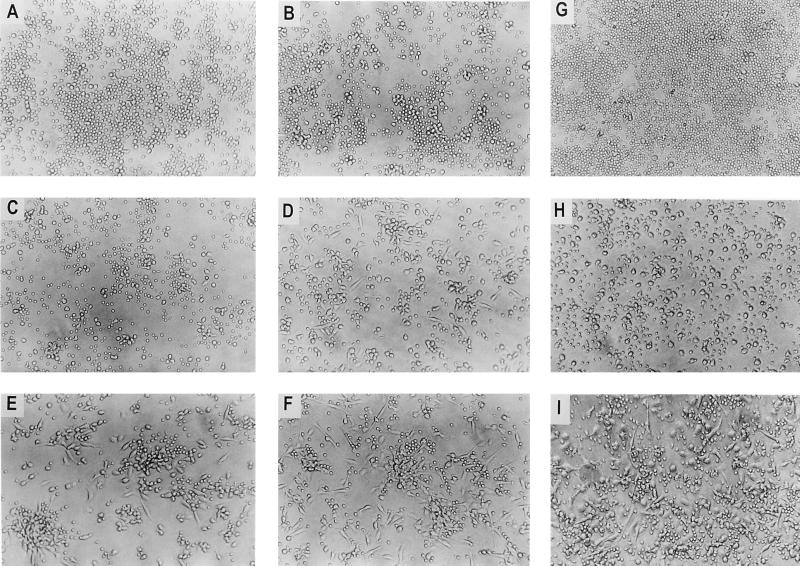
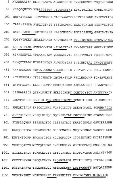
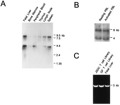
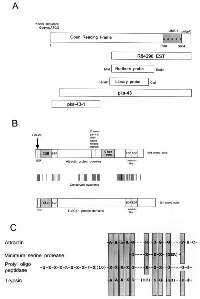
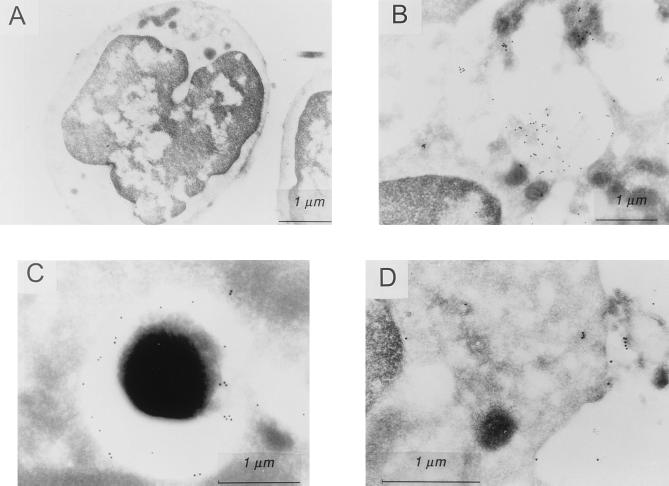
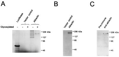
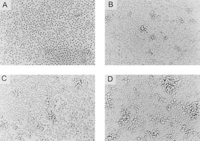
Attractin is a normal human serum glycoprotein of 175 kDa that is rapidly expressed on activated T cells and released extracellularly after 48-72 hr. We have cloned attractin and find that, as in its natural serum form, it mediates the spreading of monocytes that become the focus for the clustering of nonproliferating T lymphocytes. There are two mRNA species with hematopoietic tissue-specific expression that code for a 134-kDa protein with a putative serine protease catalytic serine, four EGF-like motifs, a CUB domain, a C type lectin domain, and a domain homologous with the ligand-binding region of the common gamma cytokine chain. Except for the latter two domains, the overall structure shares high homology with the Caenorhabditis elegans F33C8.1 protein, suggesting that attractin has evolved new domains and functions in parallel with the development of cell-mediated immunity.


Figures

Figure 1
Figure 1
Purified natural attractin mediates monocyte spreading and T cell clustering after 48 hr. (A) Control, no addition; (B) 0.5 μg/ml; (C) 1 μg/ml; (D) 2 μg/ml; (E) 5 μg/ml; (F) 10 μg/ml; Attractin at 10 μg/ml was added for 48 hr to PBMC, separated into E+ T lymphocytes (G), E monocytes/B cells (H), and to T lymphocytes remixed with monocytes/B cells (I).
Figure 2
Figure 2
Peptide sequence of attractin. The sequences identified by N-terminal sequencing of tryptic and chymotryptic peptides are underlined.
Figure 3
Figure 3
Attractin mRNA expression. (A) Multiple tissue mRNA Northern blot. (B) Resting and PHA-activated PBMC total RNA. (C) PCR amplification of a 3164-bp fragment (from 3594 bp) of attractin from three independent cDNA libraries.
Figure 4
Figure 4
Organization of attractin cDNA and peptide domains. (A) Attractin cDNA: the uppercase bases at the origin represent bases satisfying the Kozak consensus. The asterisks indicate in-frame stop codons. (B) Comparison of attractin protein domains and motifs with C. elegans F33C8.1 protein, the horizontal bars depict the position of cysteines shared by both sequences. (C) Comparison of the putative catalytic serine motif of attractin with the catalytic serine motifs of other serine proteases. The shaded box indicates agreement with the consensus, the # or exclusion from the shaded boxes indicates conflict, and the X indicates satisfaction by any amino acid. The parentheses enclose amino acids any of which would satisfy the consensus.
Figure 5
Figure 5
Intracellular localization of glycosylated attractin. Immunogold electron microscopy using rabbit polyclonal antiattractin. (A) Resting T lymphocyte with no evidence of attractin expression. (B) In T cells activated for 48 hr with PHA, attractin is expressed in large vacuoles. (C) Attractin often localizes in vesicles with electron dense core. (D) Vesicles containing attractin break open at the cell surface releasing attractin.
Figure 6
Figure 6
Expression of recombinant attractin and immunoprecipitation by antibody against natural attractin. (A) In vitro transcription/translation of attractin in absence or presence of glycosyl transferases. (B) Western blotting with anti-myc of lysates of 293T cells transiently transfected with pSecTag2B-attractin or pSecTag2B vector control. (C) Attractin was precipitated with preimmune antibody or antiattractin antibody from lysates of CHO cells stably transfected with pSecTag2B-attractin.
Figure 7
Figure 7
Recombinant attractin mediates monocyte/macrophage spreading and T cell clustering. Resting PBMC were incubated with increasing concentrations of purified recombinant attractin for 48 hr. (A) Control, no addition; (B) 1 μg/ml; (C) 2 μg/ml; (D) 5 μg/ml.

References

    1. Shimizu Y, Shaw S. FASEB J. 1991;5:2292–2299. - PubMed
    1. Gilat D, Cahalon L, Hershkoviz R, Lider O. Immunol Today. 1996;17:16–20. - PubMed
    1. Hauzenberger D, Klominek J, Bergstrom S E, Sundqvist K G. Crit Rev Immunol. 1995;15:285–316. - PubMed
    1. Masuyama J, Berman J S, Cruikshank W W, Morimoto C, Center D M. J Immunol. 1992;148:1367–1374. - PubMed
    1. Brezinschek R I, Lipsky P E, Galea P, Vita R, Oppenheimer-Marks N. J Immunol. 1995;154:3062–3077. - PubMed

Publication types

MeSH terms

Associated data

LinkOut - more resources