. 1998 Sep 15;95(19):11371-6.
doi: 10.1073/pnas.95.19.11371.

Conditional lineage ablation to model human diseases

Affiliations

Conditional lineage ablation to model human diseases

P Lee et al. Proc Natl Acad Sci U S A. .

Abstract

Cell loss contributes to the pathogenesis of many inherited and acquired human diseases. We have developed a system to conditionally ablate cells of any lineage and developmental stage in the mouse by regulated expression of the diphtheria toxin A (DTA) gene by using tetracycline-responsive promoters. As an example of this approach, we targeted expression of DTA to the hearts of adult mice to model structural abnormalities commonly observed in human cardiomyopathies. Induction of DTA expression resulted in cell loss, fibrosis, and chamber dilatation. As in many human cardiomyopathies, transgenic mice developed spontaneous arrhythmias in vivo, and programmed electrical stimulation of isolated-perfused transgenic hearts demonstrated a strikingly high incidence of spontaneous and inducible ventricular tachycardia. Affected mice showed marked perturbations of cardiac gap junction channel expression and localization, including a subset with disorganized epicardial activation patterns as revealed by optical action potential mapping. These studies provide important insights into mechanisms of arrhythmogenesis and suggest that conditional lineage ablation may have wide applicability for studies of disease pathogenesis.


Figures

Figure 1
Figure 1
(A) Kaplan–Meier curve. Matings were carried out between single transgenic MHCαtTA and tet-DTA strains. Tetracycline was administered throughout gestation and withdrawn at varying times after birth. Survival of double-transgenic offspring from the time of tetracycline withdrawal was determined. Median survival was 32 days (n = 19 binary mice). (B) Survival as a function of withdrawal age. No relationship between age at the time of tetracycline withdrawal and subsequent length of survival was observed (r2 = 0.02).
Figure 2
Figure 2
Histological analysis. Hearts were fixed in formalin and paraffin sections prepared and stained with either hematoxylin and eosin (A and B) or trichrome (CF) to highlight the interstitial fibrosis. Representative low-power views from a control heart (A) and individual binary transgenic hearts (BD). Note the chamber dilatation (B and C) and marked variability in the extent of fibrosis (C and D) in binary hearts. Higher power views demonstrate the presence of scant residual myocytes in the most severe lesions (E and F). Scales are indicated. A and B are montages.
Figure 3
Figure 3
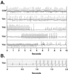
Telemetric recordings. (A) Representative rhythm strips from nontransgenic (control) and binary transgenic mice (TG1–TG4). Abnormalities including atrial fibrillation (TG1), pauses (TG2), and runs of ventricular tachycardia (TG3 and TG4) were observed. (B) Replotting of TG3 trace at faster chart speed reveals altered QRS morphology of ectopic ventricular beats.
Figure 4
Figure 4
Ventricular tachycardia in isolated-perfused hearts. Representative tracings of ventricular arrhythmias in binary transgenic mice. Recurrent bursts and nonsustained and sustained ventricular tachycardia were observed in individual hearts.
Figure 5
Figure 5
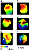
Optical action potential analysis. Isolated-perfused hearts were studied as described. Representative images showing normal anisotropic conduction observed in all control hearts (A) and most binary transgenics (B). In three transgenic mice, varying degrees of disorganized epicardial activation were observed (CE). An area of slow conduction observed during pacing (E) served as an obstacle for inducible ventricular tachycardia (F). Hearts were imaged from either the anterior (A, B, and D) or posterior (C, E, and F) surfaces.
Figure 6
Figure 6
Cx43 immunostaining. Representative confocal images of Cx43 expression in control (A) and binary transgenic hearts with mild (B), moderate (C), or severe (D) disease. Paired images show phase and immunofluorescence. In the moderately and severely diseased hearts, regions at a distance from focal fibrosis are shown. Images were acquired for equivalent times so that signal intensities could be compared directly. Substantial reduction in the intensity of junctional staining is evident in all transgenic hearts. (Original magnification was 60×.)
Figure 7
Figure 7
Western blot analysis. Companion gels either stained with Coomassie Blue to visualize total proteins loaded (A) or further processed for immunoblotting to visualize Cx43 expression (B). The distribution of total proteins in control (lanes 1–4) and transgenic (lanes 5–8) hearts was similar, but Cx43 abundance in binary transgenic hearts was significantly reduced (n = 4 for each group; P < 0.05). This experiment was repeated in triplicate.

Similar articles

Cited by

References

    1. Hetts S W. J Am Med Assoc. 1998;279:300–307. - PubMed
    1. Rinkenberger J L, Korsmeyer S J. Curr Opin Genet Dev. 1997;7:589–596. - PubMed
    1. MacLellan W R, Schneider M D. Circ Res. 1997;81:137–144. - PubMed
    1. Barbosa E R, Limongi J C, Cummings J L. Psychiatr Clin North Am. 1997;20:769–790. - PubMed
    1. Lev M. Cardiovasc Clin. 1972;4:159–186. - PubMed

Publication types

MeSH terms