. 1998 Sep 15;95(19):11406-11.
doi: 10.1073/pnas.95.19.11406.

Hippocampal morphometry in schizophrenia by high dimensional brain mapping

Affiliations

Hippocampal morphometry in schizophrenia by high dimensional brain mapping

J G Csernansky et al. Proc Natl Acad Sci U S A. .

Abstract

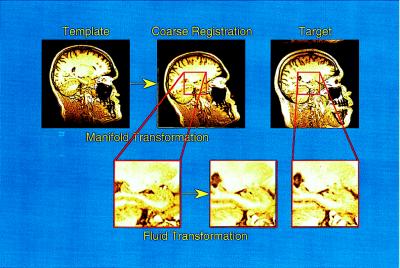
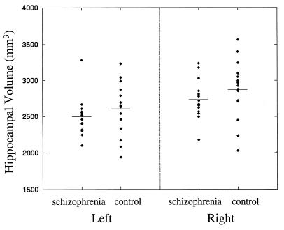
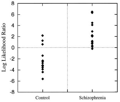
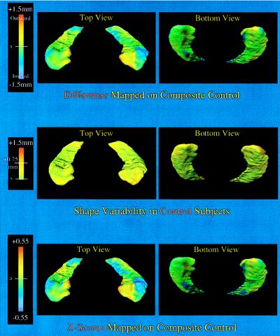
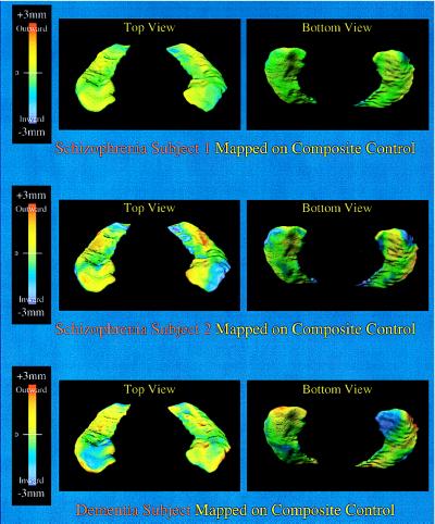
Theories of the pathophysiology of schizophrenia have implicated the hippocampus, but controversy remains regarding hippocampal abnormalities in patients with schizophrenia. In vivo studies of hippocampal anatomy using high resolution magnetic resonance scanning and manual methods for volumetric measurement have yielded inconclusive results, perhaps because of the normal variability in hippocampal volume and the error involved in manual measurement techniques. To resolve this controversy, high dimensional transformations of a computerized brain template were used to compare hippocampal volumes and shape characteristics in 15 matched pairs of schizophrenia and control subjects. The transformations were derived from principles of general pattern matching and were constrained according to the physical properties of fluids. The analysis and comparison of hippocampal shapes based on these transformations were far superior to the comparison of hippocampal volumes or other global indices of hippocampal anatomy in showing a statistically significant difference between the two groups. In the schizophrenia subjects, hippocampal shape deformations were found to be localized to subregions of the structure that send projections to prefrontal cortex. The results of this study demonstrate that abnormalities of hippocampal anatomy occur in schizophrenia and support current hypotheses that schizophrenia involves a disturbance of hippocampal-prefrontal connections. These results also show that comparisons of neuroanatomical shapes can be more informative than volume comparisons for identifying individuals with neuropsychiatric diseases, such as schizophrenia.


Figures

Figure 1
Figure 1
High dimensional transformations proceeded in a two-step process. The template was produced by using a MR scan from a healthy control subject, and information about the boundaries of the hippocampus was placed within this template by experts by using manual outlining methods. Landmarks to indicate approximate brain and hippocampal boundaries were placed in the template and each of the target scans. The first step in the transformation was guided by these landmarks and roughly oriented the template to the target scans. The second step of the transformation, performed in blocks of tissue containing the left and right hippocampus, was driven by individual voxel gray scale values but was constrained by fluid physical properties.
Figure 2
Figure 2
Hippocampal volumes were estimated by calculating the voxels enclosed by hippocampal surfaces that had been carried along through the high dimensional transformations. A comparison of the volumes in the two groups was not statistically significant (see text).
Figure 3
Figure 3
A comparison of individual log likelihood ratio values in the two groups showed a strong statistically significant difference (see text). The values shown were derived from the optimal linear combination of eigenvectors (i.e., 1, 3, 4, 6, 10, and 15), determined by a stepwise process. When we used this method of comparison, 12 of 15 subjects in each group were classified correctly.
Figure 4
Figure 4
The hippocampal shapes shown represent the composite hippocampal surfaces in the healthy controls. (Top) The degree of displacement of these surfaces (in millimeters) perpendicular to the plane of the structure and relative to the control composite is indicated by a flame scale. The lateral aspect of the head of the hippocampus and the medial aspect of the body, where the subiculum is found, showed localized shape deformities in the schizophrenia subjects. (Middle) The pattern of shape variability in controls is shown [SD at each surface point calculated by using the optimal linear combination of six eigenfuncions (i.e., 1, 3, 4, 6, 10, 15). More than 95% of surface points had a SD <0.75 mm. (Bottom) A map of z-score values is shown to examine the shape deformities in the schizophrenia subjects while accounting for general variability in hippocampal shape. In contrast to the normal pattern of shape variability, shape deformities found in the schizophrenia subjects were highly localized.
Figure 5
Figure 5
(Top and Middle) Hippocampal shape deformities in two individuals with schizophrenia with the largest log likelihood ratio values (see Fig. 3) are shown and can be compared with those found in an individual with mild dementia of the Alzheimer type and whose hippocampal volumes were similar to the schizophrenia subjects (Bottom, left hippocampal volume = 2,581 mm3, right hippocampal volume = 2448 mm3). The displacement of these surfaces (in millimeters) perpendicular to the plane of the structure and relative to the control composite is indicated by a flame scale. Shape deformities in the dementia subject were distributed differently than those found in the two schizophrenia subjects and did not specifically involve the lateral aspect of the head of the hippocampus.

Similar articles

Cited by

References

    1. Csernansky J G, Murphy G M, Faustman W O. Biol Psychiatry. 1991;30:383–400. - PubMed
    1. Roberts G W. Trends Neurosci. 1990;13:207–211. - PubMed
    1. Saykin A J, Shtasel D L, Gur R E, Kester D B, Mozley L H, Stafiniak P, Gur R C. Arch Gen Psych. 1994;51:124–131. - PubMed
    1. Falkai P, Bogerts B. Eur Arch Psychiatr Neurol Sci. 1986;236:154–161. - PubMed
    1. Jeste D V, Lohr J B. Arch Gen Psychiatry. 1989;46:1019–1024. - PubMed

Publication types

MeSH terms