. 1998 Sep 15;95(19):11465-70.
doi: 10.1073/pnas.95.19.11465.

Activation of the genetically defined m1 muscarinic receptor potentiates N-methyl-D-aspartate (NMDA) receptor currents in hippocampal pyramidal cells

Affiliations

Activation of the genetically defined m1 muscarinic receptor potentiates N-methyl-D-aspartate (NMDA) receptor currents in hippocampal pyramidal cells

M J Marino et al. Proc Natl Acad Sci U S A. .

Abstract

Evidence suggests that cholinergic input to the hippocampus plays an important role in learning and memory and that degeneration of cholinergic terminals in the hippocampus may contribute to the memory loss associated with Alzheimer's disease. One of the more prominent effects of cholinergic agonists on hippocampal physiology is the potentiation of N-methyl-D-aspartate (NMDA)-receptor currents by muscarinic agonists. Here, we employ traditional pharmacological reagents as well as m1-toxin, an m1 antagonist with unprecedented selectivity, to demonstrate that this potentiation of NMDA-receptor currents in hippocampal CA1 pyramidal cells is mediated by the genetically defined m1 muscarinic receptor. Furthermore, we demonstrate the colocalization of the m1 muscarinic receptor and the NR1a NMDA receptor subunit at the electron microscopic level, indicating a spatial relationship that would allow for physiological interactions between these two receptors. This work demonstrates that the m1-muscarinic receptor gene product modulates excitatory synaptic transmission, and it has important implications in the study of learning and memory as well as the design of drugs to treat neurodegenerative diseases such as Alzheimer's.


Figures
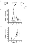
Figure 1
Figure 1
Activation of muscarinic receptors potentiates NMDA-receptor currents in CA1 pyramidal cells. Bath application of 10 μM CCh induces a marked potentiation of currents evoked by NMDA application. (A) Single traces obtained before CCh application (Pre-Drug), at the peak of CCh-induced potentiation (10 μM CCh), and after a 5-min washout. (B) The time course of CCh-induced potentiation of NMDAR currents demonstrates that this effect quickly desensitizes. Data represent mean ± SEM of five cells. (C) The potentiating effect of CCh is dose-dependent in the range of 1–100 μM. Data represent mean ± SEM of peak potentiation with three cells for each data point.
Figure 2
Figure 2
The effect of mAChR antagonists on the CCh-induced potentiation of NMDA-receptor currents. Data presented show the effect of CCh (10 μM) on peak NMDAR currents in the absence of antagonists (Control) or in the presence of the nonspecific mAChR antagonist atropine (1 μM), the m1-selective antagonist pirenzepine (75nM), or the m4-selective antagonist PD 102807 (250 nM). Data are presented as percentage of potentiation (mean ± SEM) of NMDAR current (∗, P < 0.05; n = 5 for each condition).
Figure 3
Figure 3
m1-Toxin specifically inhibits the CCh-induced potentiation of NMDA-receptor currents. (A) Time course of CCh-induced potentiation of NMDAR current in a representative cell pretreated with m1-toxin (○) and a control cell treated in the same fashion but without m1-toxin (•). (B) Mean (±SEM) data demonstrating that m1-toxin specifically blocks the potentiating effect of CCh but has no effect on the response to 1S,3R-ACPD. (∗, P < 0.05; n = 4–5 for each condition).
Figure 4
Figure 4
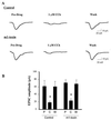
Pretreatment with m1-toxin does not inhibit the CCh-induced reduction of evoked EPSCs. (A) Single traces demonstrating that 1 μM CCh inhibits evoked EPSCs in both control and m1-toxin-treated slices. (B) Mean (±SEM) data showing that CCh produces a significant inhibition of evoked EPSCs in both control and m1-toxin-treated slices (∗, P < 0.05; n = 4 for each condition). P, pre-drug; C, 1 μM CCh; W, washout.
Figure 5
Figure 5
The m1-muscarinic receptor colocalizes with the NMDA NR1 subunit. Double-labeling electron microscopic immunocytochemistry was used to identify immunoreactivity for the m1 mAChR (visualized with silver-enhanced immunogold, electron-dense round particles) and the NR1 subunit of the NMDAR protein (visualized with DAB; a diffuse floccular reaction product). (A) A pyramidal cell soma (pc**) that contains immunoreactivity for m1 and NR1. Note the presence of DAB (dark diffuse-reaction product filling cytoplasm, and immunogold particles present in the cytoplasm and lining the membrane highlighted with small arrows in AC). Also pictured is a large proximal dendrite (d**) double-labeled with both m1 and NMDAR immunoreactivity. n, nucleus. (B) A large proximal dendrite (d**) that contains immunoreactivity for both the m1 and NMDARs. A dendritic spine (s*) containing NMDA immunoreactivity is seen extending off the proximal dendrite that is receiving an asymmetric (excitatory) synapse (arrowhead) from an unlabeled synaptic terminal (t). An unlabeled dendrite (d) is also pictured. (C) Several proximal and distal dendrites that are double-labeled for m1 and NMDAR proteins (d**) are shown. The two dendrites at the top of the panel are smaller distal dendrites, while the one at the bottom is a larger proximal dendrite. The distal dendrite on the top left can be seen receiving an asymmetric synapse (arrowhead) from an unlabeled terminal (t). Additionally, a dendritic spine (s*) containing immunoreactivity for the m1 receptor is shown extending off of the proximal dendrite. [Bar = 1.2 μm (A); 465 nm (B); 440 nm (C).]

Similar articles

Cited by

References

    1. Brown T H, Zador A M. In: The Synaptic Organization of the Brain. Shepherd G M, editor. New York: Oxford Univ. Press; 1990. pp. 346–388.
    1. Drachman D A, Leavitt J L. Arch Neurol. 1974;30:113–121. - PubMed
    1. Bartus R T, Dean D L I, Beer B, Lippa A S. Science. 1982;217:783–790. - PubMed
    1. Dekker A J A M, Connor D J, Thal L J. Neurosci Biobehav Rev. 1991;15:299–317. - PubMed
    1. Fibiger H C. Trends Neurosci. 1991;14:220–223. - PubMed

Publication types

MeSH terms

LinkOut - more resources