. 1998 Sep;113(3):367-72.
doi: 10.1046/j.1365-2249.1998.00676.x.

Expression, characterization, processing and immunogenicity of an insulin-dependent diabetes mellitus autoantigen, IA-2, in Sf-9 cells

Affiliations

Expression, characterization, processing and immunogenicity of an insulin-dependent diabetes mellitus autoantigen, IA-2, in Sf-9 cells

H Xie et al. Clin Exp Immunol. 1998 Sep.

Abstract

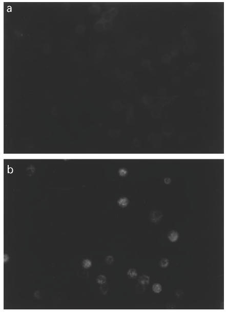
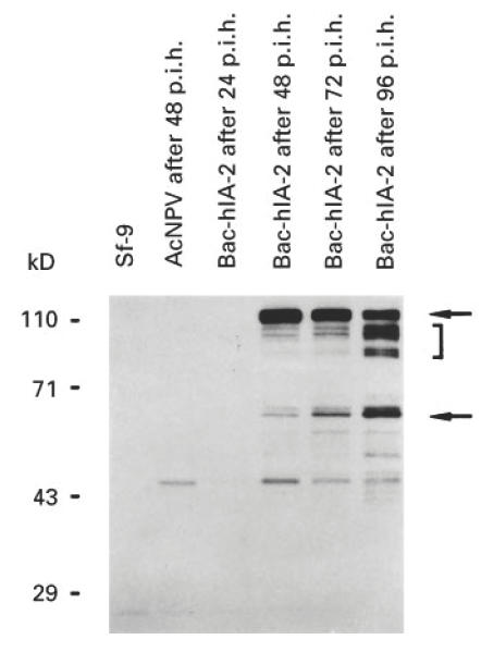
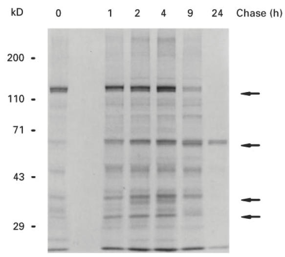
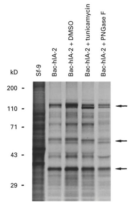
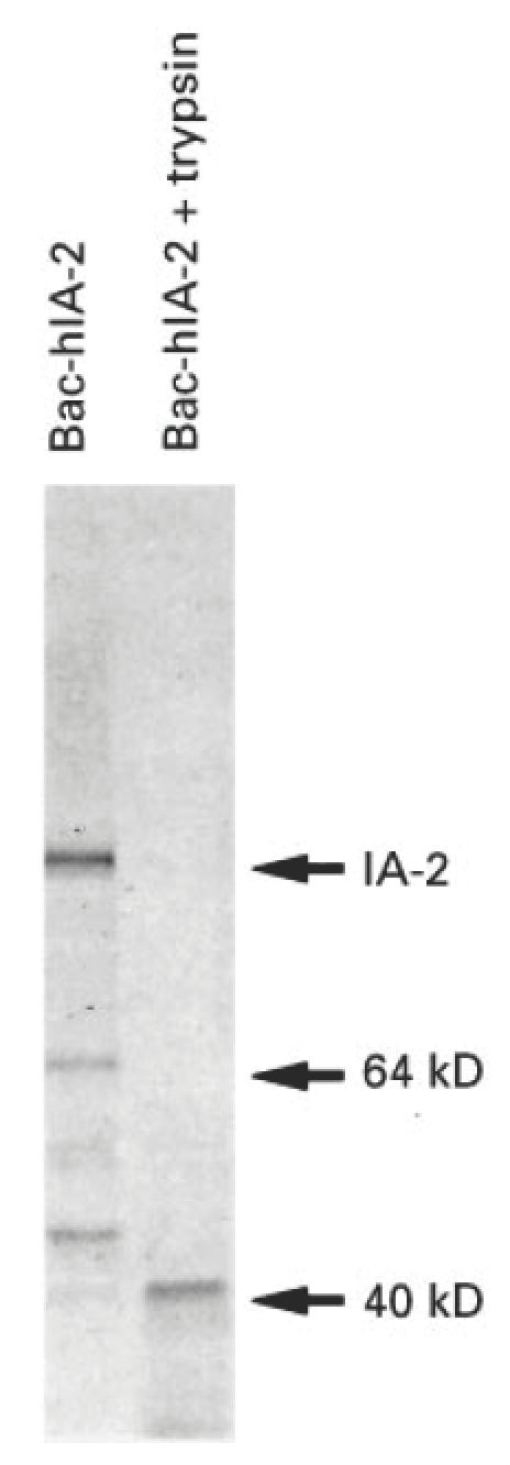
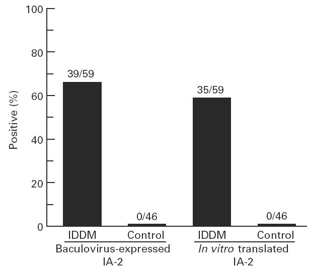
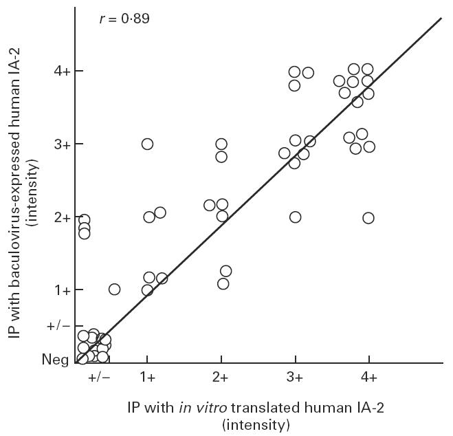
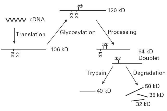
Autoantibodies to a 64-kD protein and a 40-kD tryptic fragment from pancreatic islets have been detected at high frequency in the sera of patients with insulin-dependent diabetes mellitus (IDDM). IA-2, a newly isolated transmembrane protein tyrosine phosphatase, is a major islet cell autoantigen in IDDM and the precursor of a 40-kD tryptic fragment. To express large quantities of recombinant IA-2 protein and analyse post-translational modifications we expressed full-length human IA-2 in baculovirus-infected Sf-9 cells. IA-2 expression was analysed by Western blot and by immunoprecipitation of 35S-methionine-radiolabelled proteins with rabbit antisera or IDDM sera. A 120-kD IA-2 protein was detected during the early, but not the late, phase of the infection. Pulse-chase experiments showed that the 120-kD protein was processed into fragments of 64 kD and smaller fragments of approximately 50 kD, 38 kD and 32 kD. The 64-kD fragment appeared as a doublet. Tunicamycin and PNGase F treatment down-shifted the 120-kD protein and the 64-kD doublet into lower molecular weight bands, suggesting that both were glycosylated. Trypsin treatment converted the 120-kD protein and the 64-kD doublet into a 40-kD fragment. Baculovirus-expressed IA-2 was as sensitive or slightly more sensitive than in vitro translated IA-2 in detecting autoantibodies to IA-2: 66% of sera from newly diagnosed IDDM patients reacted with baculovirus-expressed IA-2 compared with 59% of the same sera which reacted with in vitro translated IA-2. It is concluded that baculovirus-expressed IA-2 is a good source of autoantigen and that a number of lower molecular weight fragments with which IDDM autoantibodies react are derived from the 120-kD full-length IA-2 molecule.


Figures

Fig. 1
Fig. 1
Expression of human IA-2 protein in Bac-hIA-2-infected Sf-9 cells. Uninfected (a) and 2 day-infected (b) Sf-9 cells were stained with rabbit antisera to IA-2 intracellular domain.
Fig. 2
Fig. 2
Time course of Bac-hIA-2 expression in Sf-9 cells. At 24, 48, 72 and 96 h post-infection cellular lysates were subjected to 10% SDS–PAGE and immunoblotted with rabbit antiserum to the intracellular domain of human IA-2. The arrows point to the 120-kD full-length human IA-2 protein and the 64-kD IA-2 doublet. The bracket shows degradation products of IA-2. Wild-type baculovirus (AcNPV).
Fig. 3
Fig. 3
Molecular processing of Bac-hIA-2 protein in Sf-9 cells. Sf-9 cells were pulsed for 30 min with35S-methionine (zero time) and chased for 1, 2, 4, 9 and 24 h. Cellular lysates were then immunoprecipitated with anti-IA-2+ insulin-dependent diabetes mellitus (IDDM) sera and subjected to 10% SDS–PAGE. Arrows point to the 120-kD and 64-kD IA-2 proteins and the various fragments of IA-2 being processed during the chase
Fig. 4
Fig. 4
Effect of tunicamycin and peptide:N-glycosidase F (PNGase F) on Bac-hIA-2 protein. Sf-9 cells were treated with 0.1% DMSO or tunicamycin (10 μg/ml) before being infected with Bac-hIA-2 recombinant virus and labelled with 35S-methionine. The cell lysates then were immunoprecipitated with insulin-dependent diabetes mellitus (IDDM) sera and subjected to 10% SDS–PAGE. Immunoprecipitated proteins were treated with PNGase F (lane 5) before being subjected to gel separation. The arrows show the molecular mass and down-shift of the 120- and 64-kD proteins.
Fig. 5
Fig. 5
Trypsin treatment of Bac-hIA-2 protein. Infected Sf-9 cell lysates were radiolabelled with 35S-methionine, immunoprecipitated with insulin-dependent diabetes mellitus (IDDM) sera, treated with trypsin (50 μg/ml) for 30 min, and then subjected to 10% SDS–PAGE. The 120-, 64- and 50-kD IA-2 proteins were converted into a 40-kD tryptic fragment.
Fig. 6
Fig. 6
Immunoprecipitation of baculovirus-expressed human IA-2 with insulin-dependent diabetes mellitus (IDDM) serum. Sf-9 cells were labelled with 35S-methionine 48 h after infection with Bac-hIA-2 recombinant virus. Various amounts of labelled cell lysates (lanes 1 and 4, 40 000 ct/min; lanes 2 and 5, 80 000 ct/min; and lanes 3 and 6, 60 000 ct/min) were immunoprecipitated either with IDDM sera (lanes 1–3) or control sera (lanes 4–6) and then subjected to 10% SDS–PAGE.
Fig. 7
Fig. 7
Comparison of the reactivity of baculovirus-expressed IA-2 and in vitro translated IA-2 with insulin-dependent diabetes mellitus (IDDM) sera. Sera from 59 new-onset IDDM patients and 46 healthy controls were tested by radioimmunoprecipitation with either baculovirus-expressed IA-2 (80 000 ct/min) or in vitro translated IA-2 (100 000 ct/min).
Fig. 8
Fig. 8
Correlation between the intensity of immunoprecipitated IA-2 bands with baculovirus-expressed IA-2 and in vitro translated IA-2 (r = 0.89, P < 0.0001).
Fig. 9
Fig. 9
Post-translational modification of IA-2. Full-length IA-2 is processed by glycosylation and protease cleavage. The dibasic amino acids (KK) represent the potential cleavage sites from which the 64-kD doublet is generated. Trypsin treatment converts the 120-kD and 64-kD proteins into a 40-kD fragment.

Similar articles

Cited by

References

    1. Atkinson MA, Maclaren NK. The pathogenesis of insulin-dependent diabetes mellitus. N Engl J Med. 1994;331:1427–36. - PubMed
    1. Schatz D, Krischer J, Home G, et al. Islet cell antibodies predict insulin dependent diabetes in U.S. school age children as powerfully as in unaffected relatives. J Clin Invest. 1994;93:2403–7. - PMC - PubMed
    1. Bonifacio E, Bingley PJ, Shattock M, Dean BM, Dunger D, Gale EAM, Bottazzo GF. Quantification of islet-cell antibodies and prediction of insulin-dependent diabetes. Lancet. 1990;335:147–9. - PubMed
    1. Christie MR, Tun RY, Lo SS, et al. Antibodies to GAD and tryptic fragments of islet 64K antigen as distinct markers for development of IDDM. Studies with identical twins. Diabetes. 1992;41:782–7. - PubMed
    1. Christie MR, Genovese S, Cassidy D, Bosi E, Brown TJ, Lai M, Bonifacio E, Bottazzo GF. Antibodies to islet 37k antigen, but not to glutamate decarboxylase, discriminate rapid progression to IDDM in endocrine autoimmunity. Diabetes. 1994;43:1254–9. - PubMed