. 1998 Sep;113(3):407-14.
doi: 10.1046/j.1365-2249.1998.00664.x.

A variety of cytokines and immunologically relevant surface molecules are expressed by normal human skeletal muscle cells under proinflammatory stimuli

Affiliations

A variety of cytokines and immunologically relevant surface molecules are expressed by normal human skeletal muscle cells under proinflammatory stimuli

K Nagaraju et al. Clin Exp Immunol. 1998 Sep.

Abstract

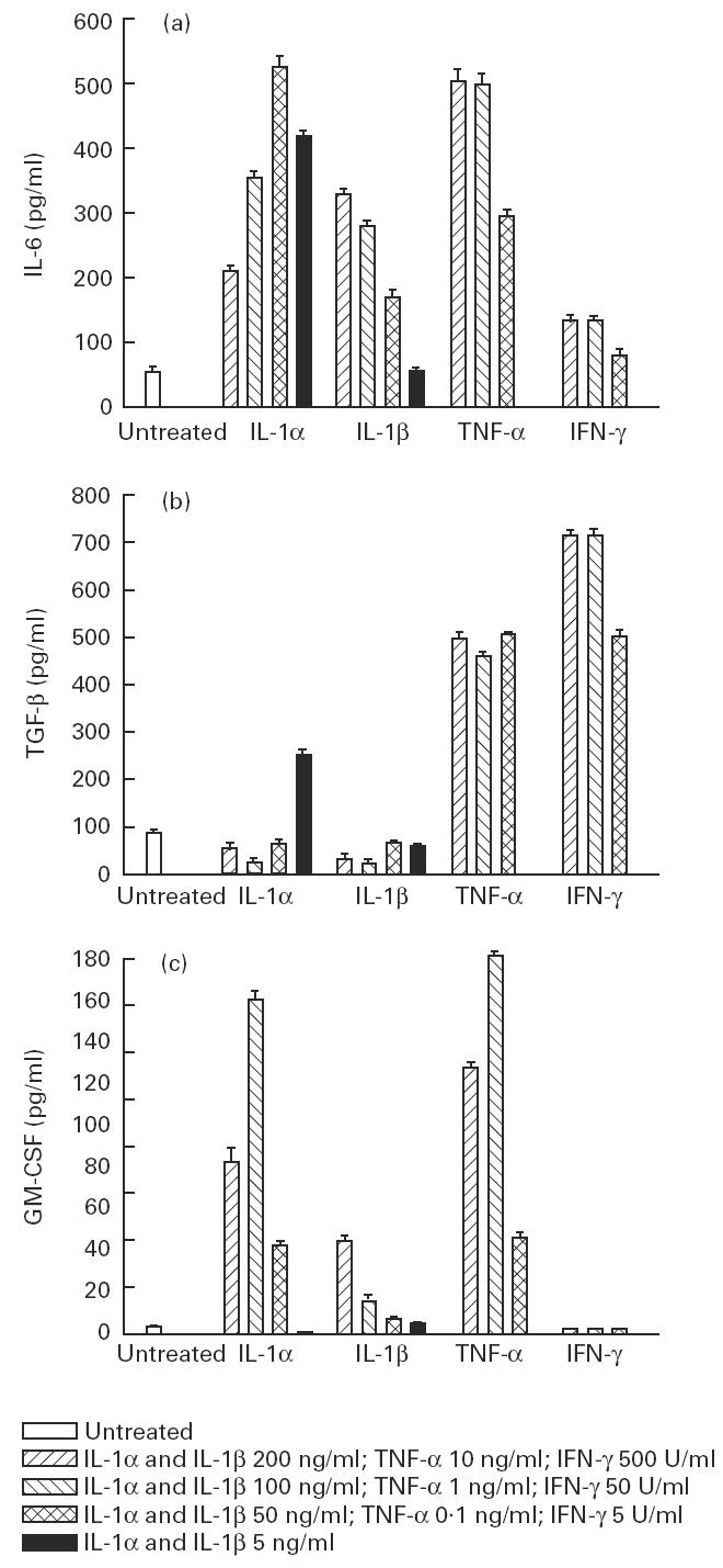
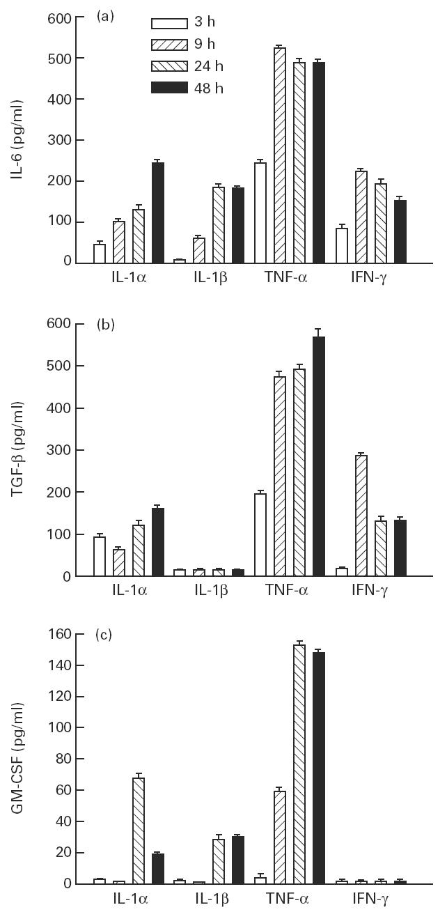
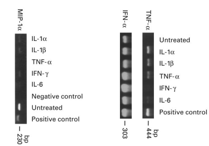
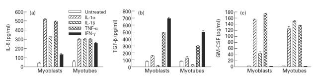
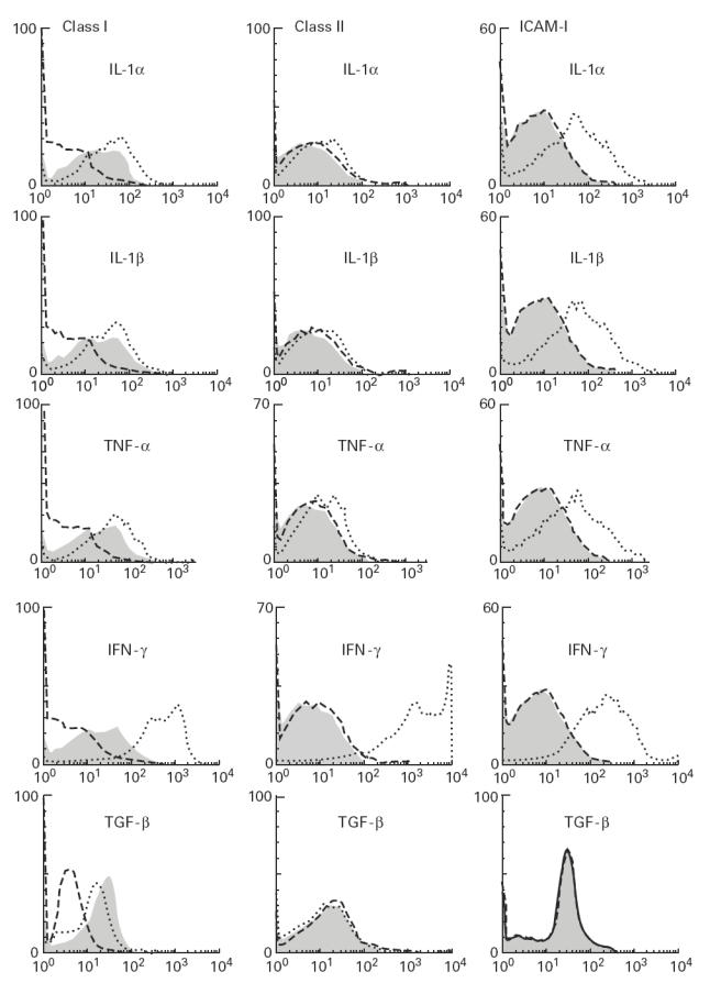
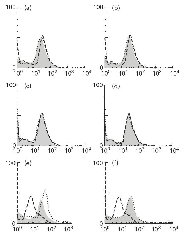
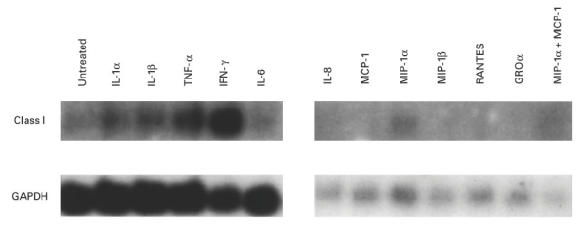
Muscle is an attractive target for gene therapy and for immunization with DNA vaccines and is also the target of immunological injury in myositis. It is important therefore to understand the immunologic capabilities of muscle cells themselves. In this study, we show that proinflammatory stimuli induce the expression of other cytokines such as IL-6, transforming growth factor-beta (TGF-beta), and granulocyte-macrophage colony-stimulating factor (GM-CSF) by muscle cells themselves, as well as the up-regulation of human leucocyte antigen (HLA) class I, class II and intercellular adhesion molecule-1 (ICAM-1). Thus, muscle cells have an inherent ability to express and respond to a variety of cytokines and chemokines. The levels of HLA class I, class II and ICAM-1 in inflamed muscle may be affected by the secreted products of the stimulation.


Figures

Fig. 1
Fig. 1
Dose effect of IL-1α, IL-1β, tumour necrosis factor-alpha (TNF-α) and IFN-γ on the production of IL-6 (a), transforming growth factor-beta (TGF-β) (b) and granulocyte-macrophage colony-stimulating factor (GM-CSF) (c) by myoblasts. The values are presented as mean (pg/ml) ± s.e.m. of three determinations in a representative experiment.
Fig. 2
Fig. 2
Effect of IL-1-α (50 ng/ml), IL-1β (100 ng/ml), tumour necrosis factor-alpha (TNF-α) (1 ng/ml) and IFN-γ (500 U/ml) on the production of IL-6 (a), transforming growth factor-beta (TGF-β) (b) and granulocyte-macrophage colony-stimulating factor (GM-CSF) (c) by myoblasts. The values are presented as mean (pg/ml) ± s.e.m. of three determinations in a representative experiment.
Fig. 3
Fig. 3
Reverse transcriptase-polymerase chain reaction (RT-PCR) analysis of the untreated and cytokine-treated muscle cultures for MIP-1α, tumour necrosis factor-alpha (TNF-α) and IFN-α expression.
Fig. 4
Fig. 4
Comparison of the levels of synthesis of IL-6 (a), transforming growth factor-beta (TGF-β) (b) and granulocyte-macrophage colony-stimulating factor (GM-CSF) (c) by myoblasts and myotubes after stimulation with IL-1α (50 ng/ml), IL-1β (100 ng/ml), tumour necrosis factor-alpha (TNF-α) (1 ng/ml) and IFN-γ (50 U/ml) for 48 h. The values are presented as mean (pg/ml) ± s.e.m. of three determinations in a representative experiment.
Fig. 5
Fig. 5
Flow cytometric analysis of myoblasts for HLA class I, HLA class II and intercellular adhesion molecule-1 (ICAM-1) after stimulation with IL-1α (1 ng/ml), IL-1β (1 ng/ml), tumour necrosis factor-alpha (TNF-α) (1 ng/ml), IFN-γ (500 U/ml) and transforming growth factor-beta (TGF-β) (10 ng/ml) for 48 h. Dashed lines, shaded areas, and dotted lines represent isotype-matched controls, unstimulated myoblasts, and stimulated myoblasts, respectively.
Fig. 6
Fig. 6
Flow cytometric analysis of myoblasts for HLA class I (e,f), HLA class II (c,d) and intercellular adhesion molecule-1 (ICAM-1) (a,b) after stimulation with MIP-1α (a,c,e) (100 ng/ml) and MCP-1 (b,d,f) (100 ng/ml) for 48 h. Dashed lines, shaded areas, and dotted lines represent isotype-matched controls, untreated myoblasts, and stimulated myoblasts, respectively.
Fig. 7
Fig. 7
Northern hybridization for the detection of HLA class I on myotubes after stimulation with IL-1α (1 ng/ml), IL-1β (1 ng/ml), tumour necrosis factor-alpha (TNF-α) (1 ng/ml), IFN-γ (500 U/ml), IL-6 (10 ng/ml) and chemokines IL-8 (100 ng/ml), MCP-1 (100 ng/ml), MIP-1α (100 ng/ml), MIP-1β (100 ng/ml), RANTES (1 ng/ml), GRO-α (100 ng/ml) and combination of MIP-1α and MCP-1 for 48 h.

Similar articles

Cited by

References

    1. Streilein JW. Peripheral tolerance induction: lessons from immune privileged sites and tissues. Transplant Proc. 1996;28:2066–70. - PubMed
    1. Law PK, Goodwin TG, Fang Q, et al. Human gene therapy with myoblast transfer. Transplant Proc. 1997;29:2234–7. - PubMed
    1. Kim JJ, Bagarazzi ML, Trivedi N, et al. Engineering of in vivo immune responses to DNA immunization via codelivery of costimulatory molecule genes. Nat Biotechnol. 1997;15:641–6. - PubMed
    1. Rosenberg NL, Ringel SP, Kotzin BL. Experimental autoimmune myositis in SJL/J mice. Clin Exp Immunol. 1987;68:117–29. - PMC - PubMed
    1. Plotz PH, Miller FW. Animal models of myositis. Mt Sinai J Med (NY) 1988;55:501–5. - PubMed

Substances