. 1998 Sep;113(3):415-22.
doi: 10.1046/j.1365-2249.1998.00666.x.

FcgammaRIa-gamma-chain complexes trigger antibody-dependent cell-mediated cytotoxicity (ADCC) in CD5+ B cell/macrophage IIA1.6 cells

Affiliations

FcgammaRIa-gamma-chain complexes trigger antibody-dependent cell-mediated cytotoxicity (ADCC) in CD5+ B cell/macrophage IIA1.6 cells

M J Van Vugt et al. Clin Exp Immunol. 1998 Sep.

Abstract

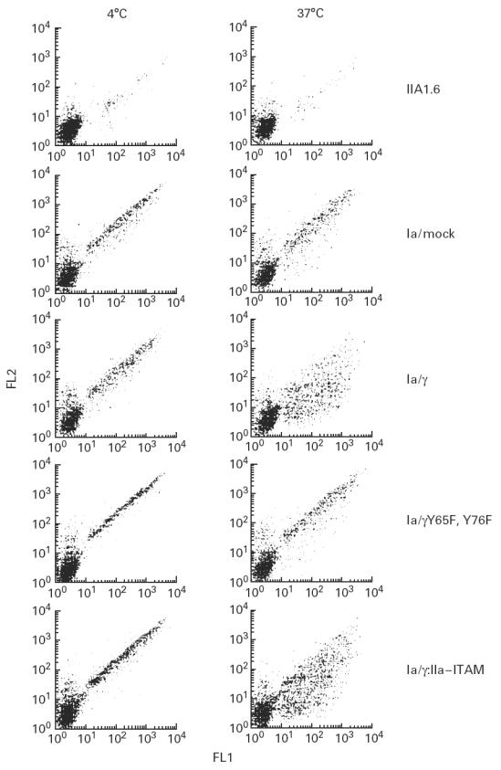
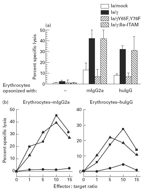
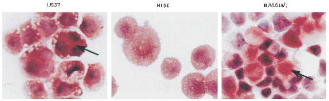
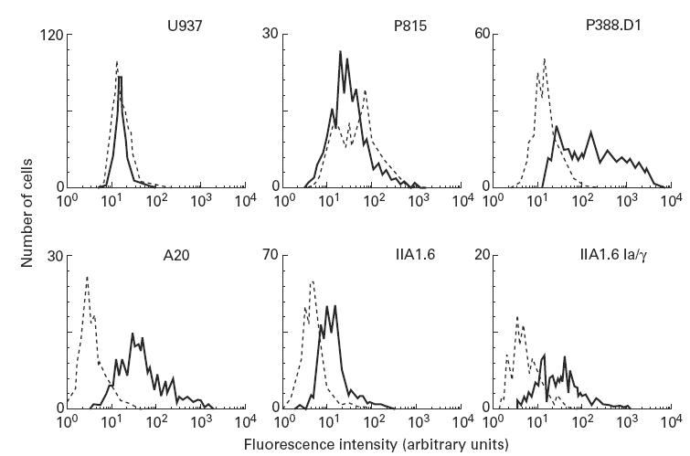
Most receptors for immunoglobulins exist as multi-subunit complexes, with unique ligand binding alpha-chains, combined with accessory signalling (gamma-, beta-, or zeta-) chains. The myeloid class I receptor for IgG (FcgammaRIa) has been shown to be dependent on the FcR gamma-chain for surface expression in vivo. In this study we assess the capacity of FcgammaRIa-gamma-chain complexes expressed in IIA1.6 cells to trigger phagocytosis and ADCC. An intact immunoreceptor tyrosine-based activation motif (ITAM) signalling motif proved essential for triggering of biological function via the FcgammaRIa receptor complex. Both the FcR gamma-chain and the FcgammaRIIa-ITAM proved active in directing phagocytosis of Staphylococcus aureus and ADCC of erythrocytes, triggered by the FcgammaRIa complex. The capacity of FcgammaRIa to trigger phagocytic and cytolytic activity by IIA1.6 cells, both considered 'professional phagocyte' functions, motivated us to re-evaluate the cell lineage and developmental stage of IIA1.6 cells. Although originally described as mouse B lymphocytes, the IIA1.6 cells proved positive for non-specific esterase activity and expressed the CD5 antigen. These combined characteristics place the IIA1.6 cells within a unique CD5+ B cell/macrophage lineage, optimally suited for cell biological analyses of phagocyte receptors.


Figures

Fig. 1
Fig. 1
Expression of FcγRIa and FcR γ-chain in IIA1.6 transfectants. Cells were incubated with FITC-labelled (CD64) MoAb 22 (solid lines), or immunofluorescence buffer alone (dotted lines). Fluorescence was recorded as arbitrary units on a logarithmic scale. Panels represent IIA1.6 cells co-transfected with FcγRIa cDNA, and with either a mock vector (a), FcR γ-chain (b), FcR γY65F,Y76F (c), or FcR γ:IIa–immunoreceptor tyrosine-based activation motif (ITAM) (d). FcR γ-chain, FcR γY65F,Y76F and FcR γ:IIa–ITAM expression was checked by reverse transcription-polymerase chain reaction (RT-PCR). The characteristic FcR γ-chain and FcR γY65F,Y76F 546 base pair, and the FcR γ:IIa–ITAM 450 base pair bands are indicated by lines. cDNA quality was confirmed by RT-PCR of HPRT. Origins of RT cDNAs are shown above the lanes (e).
Fig. 2
Fig. 2
Phagocytosis by FcγRIa-expressing IIA1.6 cells. FITC-labelled IgG-opsonized Staphylococcus aureus were incubated with either non-transfected cells, or FcγRIa complex-transfected cells for 45 min at 4°C. Cells were further incubated for 45 min either at 4°C (left lanes) or 37°C (right lanes). Remaining cell surface-bound bacteria were stained with PE-conjugated goat anti-human antibody. FITC (FL1) and PE (FL2) fluorescence of 3500 cells was quantified by flow cytometry, and dot plot diagrams are shown. Experiments were repeated at least four times yielding almost identical results.
Fig. 3
Fig. 3
(a) ADCC triggered by FcγRIa-transfected IIA1.6 cells. Lysis of erythrocytes was measured by 51Cr release in supernatants after 3 h coincubation of IIA1.6 transfectants with mouse IgG2a (mIgG2a)- or human IgG (huIgG)-opsonized erythrocytes (effector:target ratio of 10). Non-opsonized erythrocytes (−) served as a control. Data represent mean ± s.e.m. of eight individual experiments. ADCC of (mIgG2a- and huIgG)-opsonized erythrocytes by FcγRIa/γ and FcγRIa/γ:IIa–immunoreceptor tyrosine-based activation motif (ITAM)-transfected cells differed significantly from other transfectants and non-transfected IIA1.6 cells (P < 0.05). (b) Erythrocyte lysis at different effector:target ratios by FcγRIa-transfected IIA1.6 cells. IIA1.6 cells expressing either FcγRIa/mock (•), FcγRIa/γ wild type (▪) or FcγRIa/γ:IIa–ITAM (▴) were incubated at different effector:target ratios (indicated on the abscissas) with either mouse IgG2a-opsonized erythrocytes (erythrocytes–mIgG2a) or human IgG-opsonized erythrocytes (erythrocytes–huIgG). Experiments were repeated at least three times, yielding essentially identical results.
Fig. 4
Fig. 4
Non-specific esterase activity in IIA1.6 cells. U937, HL-60 and FcγRIa-transfected IIA1.6 cells were stained for non-specific esterase activity, by incubation with hexazonium pararosanilin followed by haematoxylin (see Materials and Methods). This staining method results in blue nuclei and cytoplasm of non-specific esterase-positive cells colouring brown/red (indicated by arrows). This experiment was repeated twice with identical results.
Fig. 5
Fig. 5
CD5 expression levels on human and murine cell lines. Human monocytic (U937), murine mastocytoma (P815), murine B cell-derived macrophage (P388.D1), murine lymphoid (A20), murine Fc receptor-deficient (IIA1.6), and FcγRIa–γ-chain transfected IIA1.6 cells were stained for murine CD5. Cells were incubated for 30 min with a biotin-labelled CD5 antibody, and subsequently with streptavidin–PE for 30 min (solid lines). A biotin-labelled isotype-matched antibody specific for a mouse mast cell marker (4D11) served as a negative control (dotted line). Experiments were performed at least three times, giving essentially identical results.

Similar articles

Cited by

References

    1. Van de Winkel JGJ, Capel PJA. In: Human IgG Fc receptors. Austin Texas, Landes R.G., editors. 1996.
    1. Hibbs ML, Selvaraj P, Carpen O, Springer TA, Kuster H, Jouvin M-H, Kinet J-P. Mechanisms for regulating expression of membrane isoforms of FcγRIII (CD16) Science. 1989;246:1608–11. - PubMed
    1. Blank U, Ra C, Miller L, White K, Metger H, Kinet J-P. Complete structure and expression in transfected cells of high affinity IgE receptor. Nature. 1989;337:187–9. - PubMed
    1. Van Vugt MJ, Heijnen Iafm, Capel PJA, Park SY, Ra C, Saito T, Verbeek JS, Van de Winkel JGJ. FcR γ-chain is essential for both surface expression and function of human FcγRI (CD64) in vivo. Blood. 1996;87:3593–9. - PubMed
    1. Isashi Y, Yamashita T, Nagasawa S, Murakami M, Uede T. Molecular cloning and characterization of guinea pig FcγRIII: expression but not function is independent of the γ chain of FcɛRI. Int Immunol. 1996;8:1335–46. - PubMed

Publication types