

Interaction of murine macrophage-membrane proteins with components of the pathogenic fungus Histoplasma capsulatum
- PMID: 9737672
- PMCID: PMC1905054
- DOI: 10.1046/j.1365-2249.1998.00656.x
Interaction of murine macrophage-membrane proteins with components of the pathogenic fungus Histoplasma capsulatum
Abstract
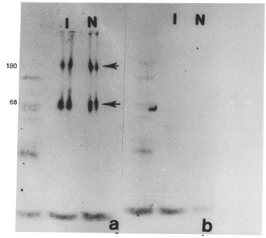
The interaction of macrophage-membrane proteins and histoplasmin, a crude antigen of the pathogenic fungus Histoplasma capsulatum, was studied using murine peritoneal macrophages. Membrane proteins were purified via membrane attachment to polycationic beads and solubilized in Tris-HCl/SDS/DTT/glycerol for protein extraction; afterwards they were adsorbed or not with H. capsulatum yeast or lectin binding-enriched by affinity chromatography. Membrane proteins and histoplasmin interactions were detected by ELISA and immunoblotting assays using anti-H. capsulatum human or mouse serum and biotinylated goat anti-human or anti-mouse IgG/streptavidin-peroxidase system to reveal the interaction. Results indicate that macrophage-membrane proteins and histoplasmin components interact in a dose-dependent reaction, and adsorption of macrophage-membrane proteins by yeast cells induces a critical decrease in the interaction. Macrophage-membrane glycoproteins with terminal D-galactosyl residues, purified by chromatography with Abrus precatorius lectin, bound to histoplasmin; and two bands of 68kD and 180kD of transferred membrane protein samples interacted with histoplasmin components, as revealed by immunoblot assays. Specificity for beta-galactoside residues on the macrophage-membrane was confirmed by galactose inhibition of the interaction between macrophage-membrane proteins and histoplasmin components, in competitive ELISA using sugars, as well as by enzymatic cleavage of the galactoside residues.
Figures


References
-
- Bullock WE. Interactions between human phagocytic cells and Histoplasma capsulatum. Arch Med Res. 1993;24:219–23. - PubMed
-
- Newman SL, Gootee L, Morris R, Bullock WE. Digestion of Histoplasma capsulatum yeasts by human macrophages. J Immunol. 1992;149:574–80. - PubMed
-
- Falkow S, Isberg RR, Portnoy DA. The interaction of bacteria with mammalian cells. Annu Rev Cell Biol. 1992;8:333–63. - PubMed
-
- Schnur RA, Newman SL. The respiratory burst response to Histoplasma capsulatum by human neutrophils. Evidence for intracellular trapping of superoxide anion. J Immunol. 1990;144:4765–72. - PubMed
Publication types
MeSH terms
Substances
LinkOut - more resources
Full Text Sources
