. 1998 Oct;36(10):2810-6.
doi: 10.1128/JCM.36.10.2810-2816.1998.

Improved amplification of microbial DNA from blood cultures by removal of the PCR inhibitor sodium polyanetholesulfonate

Affiliations

Improved amplification of microbial DNA from blood cultures by removal of the PCR inhibitor sodium polyanetholesulfonate

D N Fredricks et al. J Clin Microbiol. 1998 Oct.

Abstract

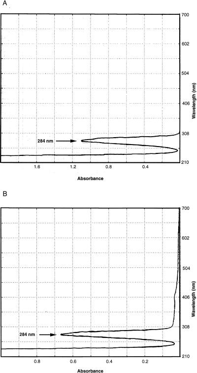
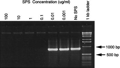
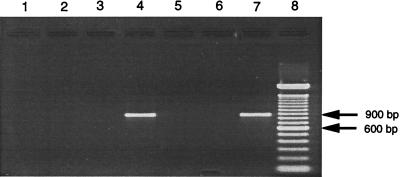
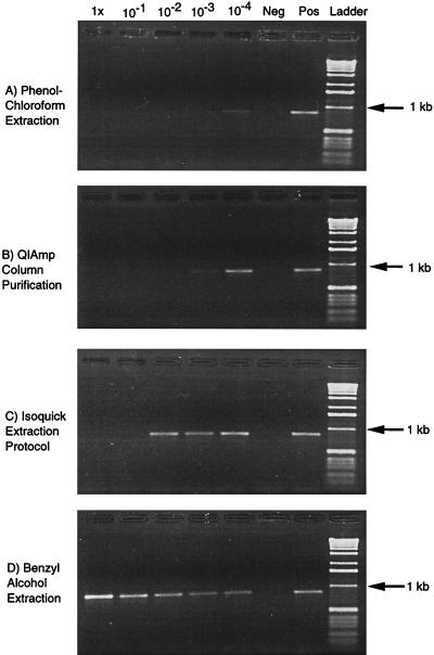
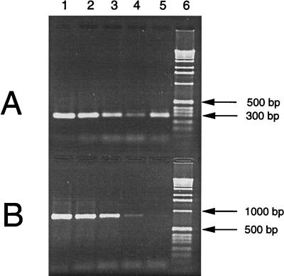
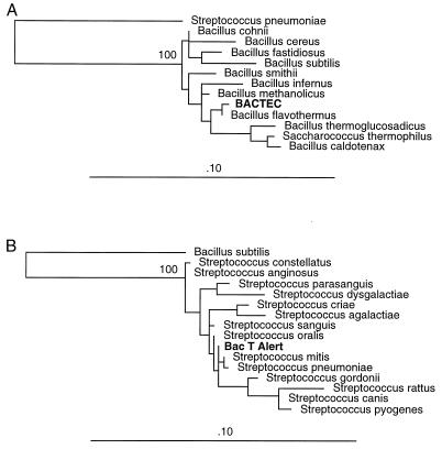
Molecular methods are increasingly used to identify microbes in clinical samples. A common technical problem with PCR is failed amplification due to the presence of PCR inhibitors. Initial attempts at amplification of the bacterial 16S rRNA gene from inoculated blood culture media failed for this reason. The inhibitor persisted, despite numerous attempts to purify the DNA, and was identified as sodium polyanetholesulfonate (SPS), a common additive to blood culture media. Like DNA, SPS is a high-molecular-weight polyanion that is soluble in water but insoluble in alcohol. Accordingly, SPS tends to copurify with DNA. An extraction method was designed for purification of DNA from blood culture media and removal of SPS. Blood culture media containing human blood and spiked with Escherichia coli was subjected to an organic extraction procedure with benzyl alcohol, and removal of SPS was documented spectrophotometrically. Successful amplification of the extracted E. coli 16S rRNA gene was achieved by adding 5 microliter of undiluted processed sample DNA to a 50-microliter PCR mixture. When using other purification methods, the inhibitory effect of SPS could be overcome only by dilution of these samples. By our extraction technique, even uninoculated blood culture media were found to contain bacterial DNA when they were subjected to broad-range 16S rRNA gene consensus PCR. We conclude that the blood culture additive SPS is a potent inhibitor of PCR, is resistant to removal by traditional DNA purification methods, but can be removed by a benzyl alcohol extraction protocol that results in improved PCR performance.


Figures

FIG. 1
FIG. 1
Spectrophotometric scan. (A) SPS at 0.0135% dissolved in water. (B) BacT Alert blood culture medium (0.1 ml) inoculated with blood was washed three times by centrifugation in 10 volumes of water (method F) and was then processed by the QIAmp blood digestion and DNA purification protocol (method B). Water eluate from the silica column, which normally contains purified DNA, was scanned.
FIG. 2
FIG. 2
Effect of SPS concentration on PCR. Agarose gel electrophoresis of PCR products amplified from B. pertussis target DNA with conserved bacterial 16S rRNA gene primers 516F and 13R in the presence of various concentrations of SPS in each PCR reaction, as listed, including no SPS (positive control). The amplification product size is about 874 bp.
FIG. 3
FIG. 3
Amplification of bacterial DNA from blood cultures by four digestion and extraction methods. Agarose gel electrophoresis of PCR products amplified with 16S rRNA gene primers 516F and 13R. In the first four lanes, E. coli was inoculated into blood culture medium containing human blood and was processed by various digestion and purification protocols to produce target DNA for PCR. Lane 1, proteinase K digestion with phenol-chloroform extractions (method A); lane 2, Isoquick protocol (method C); lane 3, protocol with the QIAmp blood kit; (method B); lane 4, benzyl alcohol-guanidine hydrochloride extraction protocol (method D); lane 5, method D performed with sterile LB broth inoculated into blood culture medium (negative control); lane 6, method D performed with sterile LB broth added to TE buffer (negative control); lane 7, bacterial DNA-positive control; lane 8, 100-bp DNA ladder. The amplification product size is about 874 bp.
FIG. 4
FIG. 4
Amplification controls. Agarose gel electrophoresis of PCR products amplified with 16S rRNA gene primers 516F and 13R (about 874 bp). E. coli was inoculated into blood culture medium containing human blood and processed by one of the four listed digestion and purification protocols. In the first five lanes, the processed DNA was diluted in sterile, UV-irradiated water as indicated, and 5 μl of DNA target were added to a 50-μl PCR mixture along with 1 μl of additional B. pertussis DNA. Lane Neg, unprocessed, sterile water (negative control); and Lane Pos, 1 μl (79 ng) of B. pertussis DNA (positive control) subjected to PCR. A 1-kb DNA ladder is present in the last lane.
FIG. 5
FIG. 5
Comparison of two primer pairs used in PCR amplification. PCR was performed with primer pair 516F-806R (A) or 516F-13R (B), and the products were subjected to gel electrophoresis. One microliter of target was added to each 50-μl PCR mixture, as follows: 20,000 gene copies of E. coli 16S rDNA (lane 1), 2,000 gene copies (lane 2), 200 gene copies (lane 3), 20 gene copies (lane 4), DNA purified from uninoculated BacT Alert anaerobic blood culture medium by method D (lane 5), and 1-kb DNA ladder (lane 6).
FIG. 6
FIG. 6
Phylogenetic trees. The 16S rRNA gene sequence amplified from uninoculated blood culture medium was aligned with other known 16S rRNA gene sequences, and phylogenetic relationships were inferred by using a maximum likelihood algorithm. The branch length is proportional to the evolutionary distance, and the bar labeled .10 represents 0.1 estimated base changes per position. (A) Sequence in BACTEC medium with 370 masked positions. S. pneumoniae was used as an outgroup. (B) Sequence in BacT Alert medium with 554 masked positions. B. subtilis was used as an outgroup.

References

    1. Ahokas H, Erkkila M J. Interference of PCR amplification by the polyamines, spermine and spermidine. PCR Methods Appl. 1993;3:65–68. - PubMed
    1. Akane A, Matsubara K, Nakamura H, Takahashi S, Kimura K. Identification of the heme compound copurified with deoxyribonucleic acid (DNA) from bloodstains, a major inhibitor of polymerase chain reaction (PCR) amplification. J Forensic Sci. 1994;39:362–372. - PubMed
    1. Altschul S F, Gish W, Miller W, Myers E W, Lipman D J. Basic local alignment search tool. J Mol Biol. 1990;215:403–410. - PubMed
    1. Benson D A, Boguski M S, Lipman D J, Ostell J, Ouellette B F. GenBank. Nucleic Acids Res. 1998;26:1–7. - PMC - PubMed
    1. Brosius J, Dull T J, Sleeter D D, Noller H F. Gene organization and primary structure of a ribosomal RNA operon from Escherichia coli. J Mol Biol. 1981;148:107–127. - PubMed

Publication types

Associated data

LinkOut - more resources