. 1998 Oct;36(10):2990-5.
doi: 10.1128/JCM.36.10.2990-2995.1998.

Multiplex PCR for typing and subtyping influenza and respiratory syncytial viruses

Affiliations

Multiplex PCR for typing and subtyping influenza and respiratory syncytial viruses

J Stockton et al. J Clin Microbiol. 1998 Oct.

Abstract

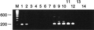
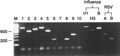
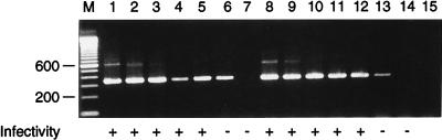
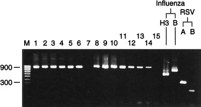
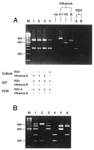
A multiplex reverse transcription (RT)-PCR method that has been developed is capable of detecting and subtyping influenza A (H1N1 and H3N2) and B viruses as well as respiratory syncytial virus (RSV) types A and B in respiratory clinical samples taken as part of a national community-based surveillance program of influenza-like illness in England and Wales. The detection of each different pathogen depended on distinguishing five amplification products of different sizes on agarose gels following RT-PCR with multiple primer sets. The multiplex RT-PCR was tested with 65 nasopharyngeal apirates from which RSV had been isolated and 237 combined nose and throat swabs from which influenza A (H1N1 and H3N2) or B virus had been detected by virus isolation, as well as 40 respiratory samples from which other viruses including cytomegalovirus, herpes simplex virus, enteroviruses, and parainfluenza viruses had been grown. For the typing and subtyping of influenza A and B viruses and RSV types A and B, the multiplex RT-PCR gave an excellent (100%) correlation with the results of conventional typing and subtyping with specific antisera. Multiplex RT-PCR can also be used to accurately detect more than one viral template in the same reaction mixture, allowing viral coinfections to be identified with the same respiratory specimen.


Figures

FIG. 1
FIG. 1
Serial 10-fold dilutions of tissue culture-grown RSV type B VS1039 were prepared in virus transport medium (starting concentration of RSV, 105 PFU/ml [lanes 1 and 8]). Each dilution was subjected to multiplex RT-PCR with all of the primer sets. Lanes 1 to 6; 10 mM Tris-HCl (pH 8.4), 1.5 mM MgCl2, 25 mM KCl, and 1.5 U of Taq in the primary reaction mixture and 0.75 U of Taq in the secondary reaction mixture; lanes 8 to 13, 10 mM Tris (pH 8.8), 3.5 mM MgCl2, 2.5 mM KCl, and 1.5 U of Taq in the primary reaction mixture and 1.5 U of Taq in the secondary reaction mixture.; lanes 7 and 14, water controls; lane M, molecular size marker. Numbers on the left are in base pairs.
FIG. 2
FIG. 2
Typing and subtyping of influenza virus and RSV in a panel of clinical specimens by multiplex RT-PCR. Lanes 1 to 6, 8, and 9, combined nose and throat swabs; lane 1, influenza A (H1N1) virus; lane 2, influenza A (H3N2) virus; lane 3, influenza B virus; lane 4, influenza A (H1N1) virus; lane 5, influenza A (H3N2) virus; lane 6, RSV type B; lane 7, virus transport medium; lane 8, RSV type B; lane 9, RSV type A; lane 10, water control. Influenza virus controls were A/Taiwan/1/86 H1N1, A/Thessaloniki/1/95 H3N2, and B/Harbin 7/94, and RSV controls were RSV type A Long and RSV type B VS1039. Amplicons were analyzed by electrophoresis on a 2.25% NuSieve agarose gel stained with ethidium bromide. Lane M, molecular size marker. Numbers on the left are in base pairs.
FIG. 3
FIG. 3
Serial 10-fold dilutions of tissue culture-grown RSV type A (Long) were prepared in virus transport medium (starting dilution, 105 PFU/ml [lane 1]). Detection of RSV PFU in 100 μl of each dilution by the infectivity assay is indicated by the plus signs. Material equivalent to 75 μl of each dilution was subjected to either multiplex RT-PCR (with all the primer sets [lanes 1 to 7]) or uniplex RT-PCR (containing only the RSV type A primer sets [lanes 8 to 14]). Lane 15, water control; lane M, molecular size marker. Numbers on the left are in base pairs.
FIG. 4
FIG. 4
Comparison of freshly prepared and stored reagents. Multiplex RT-PCR (with all the primer sets) was performed with a 10-fold dilution series of A/Taiwan/1/86 (H1N1) starting at 10−1 with both freshly prepared amplification mixtures (lanes 1 to 7) and amplification mixtures frozen for 6 months at −20°C (lanes 8 to 14). Results for influenza virus controls (A/Thessaloniki/1/95 H3N2; B/Harbin/7/94 B) and RSV controls (RSV type A Long, RSV type B VS1039) are shown. Lane 15, water control; Lane M, molecular size marker. Numbers on the left are in base pairs.
FIG. 5
FIG. 5
Simultaneous detection of multiple templates. (A) Lanes 1 to 4, sequential clinical specimens from an immunosuppressed child. Corresponding culture and direct immunofluorescence (DIF) results are shown. Sample types were bronchoalveolar lavages (lane 1), endotracheal aspirates (lane 2), and nasopharyngeal aspirates (lanes 3 and 4). Specimens in all four lanes were positive for RSV by direct immunofluorescence, the samples in lanes 1 to 3 were negative for RSV by culture, the sample in lane 4 was positive for RSV by culture, the samples in all four lanes were positive for RSV type A by PCR, the samples in all four lanes were negative for influenza virus by direct immunofluorescence, the samples in lanes 1 to 3 were positive for influenza virus by culture, the sample in lane 4 was negative for influenza virus by culture, and the samples in all four lanes were positive for influenza B virus by PCR. (B) Multiplex RT-PCR (with all primer sets) was performed with combined nose and throat swabs spiked with various combinations of influenza and RSV templates. Lanes 1 to 6 were positive (+) or negative (−) for influenza A virus H1N1, influenza A virus H3N2, influenza B virus, RSV type A, and RSV type B, as follows: lane 1, +, +, +, +, and +, respectively; lane 2, +, −, −, −, and +, respectively; lane 3, +, +, +, −, and −, respectively; lane 4, −, −, −, +, and +, respectively; lane 5, −, +, +, −, and −, respectively; lane 6, −, +, −, +, and −, respectively. (A and B) Lanes M, molecular size markers. Numbers on the left are in base pairs.

References

    1. Anderson L J, Hierholzer J C, Tsou C, Hendry R M, Fernie B F, Stone Y, McIntosh K. Antigenic characterisation of respiratory syncytial virus strains with monoclonal antibodies. J Infect Dis. 1985;163:626–633. - PubMed
    1. Axton R A, Brock D J. A single tube multiplex system for the simultaneous detection of 10 common cystic fibrosis mutations. Hum Mutat. 1995;5:260–262. - PubMed
    1. Boom R, Sol C J A, Salimans M M M, Jansen C L, Wertheim-van Dillion P M E, van der Noordaa J. Rapid and simple method for purification of nucleic acids. J Clin Microbiol. 1990;28:495–503. - PMC - PubMed
    1. Chakraverty P. Antigenic relationships between influenza B viruses. Bull W H O. 1971;45:755–766. - PMC - PubMed
    1. Chamberlain J S, Gibbs R A, Rainer J E, Nguyen P N, Caskey C T. Deletion screening of the muscular dystrophy locus via multiplex DNA amplification. Nucleic Acids Res. 1988;16:11141–11156. - PMC - PubMed

MeSH terms

Substances

LinkOut - more resources