. 1998 Oct;18(10):5670-7.
doi: 10.1128/MCB.18.10.5670.

Degradation of myogenic transcription factor MyoD by the ubiquitin pathway in vivo and in vitro: regulation by specific DNA binding

Affiliations

Degradation of myogenic transcription factor MyoD by the ubiquitin pathway in vivo and in vitro: regulation by specific DNA binding

O Abu Hatoum et al. Mol Cell Biol. 1998 Oct.

Abstract

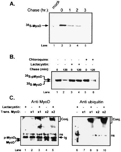
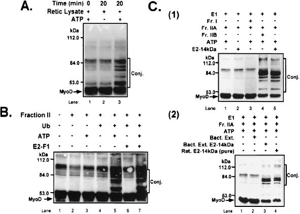
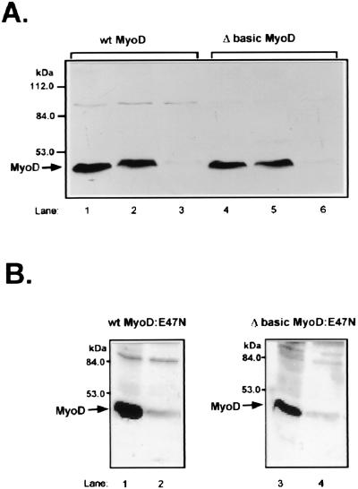
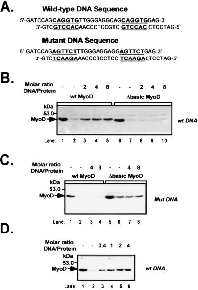
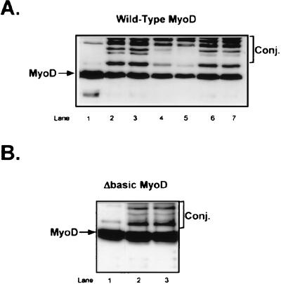
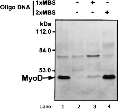
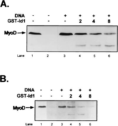
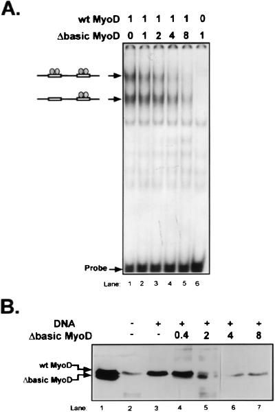
MyoD is a tissue-specific transcriptional activator that acts as a master switch for skeletal muscle differentiation. Its activity is induced during the transition from proliferating, nondifferentiated myoblasts to resting, well-differentiated myotubes. Like many other transcriptional regulators, it is a short-lived protein; however, the targeting proteolytic pathway and the underlying regulatory mechanisms involved in the process have remained obscure. It has recently been shown that many short-lived regulatory proteins are degraded by the ubiquitin system. Degradation of a protein by the ubiquitin system proceeds via two distinct and successive steps, conjugation of multiple molecules of ubiquitin to the target protein and degradation of the tagged substrate by the 26S proteasome. Here we show that MyoD is degraded by the ubiquitin system both in vivo and in vitro. In intact cells, the degradation is inhibited by lactacystin, a specific inhibitor of the 26S proteasome. Inhibition is accompanied by accumulation of high-molecular-mass MyoD-ubiquitin conjugates. In a cell-free system, the proteolytic process requires both ATP and ubiquitin and, like the in vivo process, is preceded by formation of ubiquitin conjugates of the transcription factor. Interestingly, the process is inhibited by the specific DNA sequence to which MyoD binds: conjugation and degradation of a MyoD mutant protein which lacks the DNA-binding domain are not inhibited. The inhibitory effect of the DNA requires the formation of a complex between the DNA and the MyoD protein. Id1, which inhibits the binding of MyoD complexes to DNA, abrogates the effect of DNA on stabilization of the protein.


Figures

FIG. 1
FIG. 1
MyoD is a short-lived protein, and its degradation in cells is mediated by the ubiquitin-proteasome pathway. (A) Stability of MyoD in vivo. The half-life of MyoD in Cos cells was measured in a pulse-chase experiment followed by immunoprecipitation of the labeled protein as described in Materials and Methods. (B) Sensitivity of MyoD degradation to cellular proteolysis inhibitors. Degradation of MyoD in Cos cells was monitored in a pulse-chase experiment followed by immunoprecipitation of the labeled protein in the presence of the proteasome inhibitor lactacystin or the lysosomal inhibitor chloroquine as described in Materials and Methods. (C) Ubiquitin-MyoD conjugates in cells. Cos cells were transfected with either 5 or 10 μg of MyoD expression vector. Following 1 h of incubation in the presence or absence of lactacystin, cells were disrupted and extracts were immunoprecipitated with anti-MyoD antibody and resolved by SDS-PAGE. Proteins were detected by Western blot analysis. Lanes 1 to 5, detection with anti-MyoD antibody. Lanes 6 to 10, following stripping of membrane, proteins were redetected with antiubiquitin antibody. pMyoD, phosphorylated form of MyoD; Conj., conjugates; Trans. MyoD, transfection with MyoD expression vector; ns, nonspecific cross-reacting protein; Ig, heavy chain of the Ig molecule.
FIG. 2
FIG. 2
ATP- and ubiquitin-dependent conjugation of MyoD in crude (A) and fractionated (B) reticulocyte lysate and in a system reconstituted from purified and partially purified enzymes (C). (A) ATP-dependent conjugation of MyoD. Conjugation of MyoD to ubiquitin in crude reticulocyte (Retic) lysate was monitored as described in Materials and Methods. Reaction mixtures containing ATP also contain an ATP-regenerating system. Reaction mixtures without ATP contain hexokinase and 2-deoxyglucose. Lane 1, reaction mixture with ATP incubated on ice; lanes 2 and 3, reaction mixtures incubated at 37°C. Conj., conjugates. (B) Conjugation of MyoD in fractionated reticulocyte lysate. Conjugation of MyoD in fractionated reticulocyte lysate was monitored as described in Materials and Methods. (C) Conjugation of MyoD requires E2-14kDa. Panel 1, conjugation of MyoD was monitored in reaction mixtures containing purified E1 and E2-14kDa and crude reticulocyte fraction (Fr.) I, IIA, and IIB as described in Materials and Methods. Panel 2, conjugation of MyoD was determined in the presence of control BL21 extract (Bact. Ext.), BL21 extract prepared from cells that express E2-14kDa, and E2-14kDa initially purified via ubiquitin affinity chromatography and further purified via anion-exchange chromatography to resolve the enzyme from other species of E2. E1, fraction IIA, and ATP were added as described for panel 1.
FIG. 3
FIG. 3
ATP- and ubiquitin-dependent degradation of wt and Δbasic homo- and heterodimers of MyoD with E47N in crude reticulocyte fraction II. (A) Degradation of wt and Δbasic MyoD homodimers. Proteolysis was monitored in the presence of crude reticulocyte fraction II by Western blot analysis as described in Materials and Methods. Lanes 1 and 4, reaction mixtures were incubated in the presence of ubiquitin but in the absence of ATP. Lanes 2 and 5, mixtures were incubated in the presence of ATP and in the absence of ubiquitin. Lanes 3 and 6, mixtures were incubated in the presence of ubiquitin and ATP. (B) Degradation of wt and Δbasic MyoD heterodimers with E47N. Proteolysis of MyoD-E47N heterodimers was monitored in the absence (lanes 1 and 3) or presence (lanes 2 and 4) of ATP as described in Materials and Methods and for panel A.
FIG. 4
FIG. 4
Binding of MyoD to its specific DNA recognition motif protects the protein from degradation. Degradation of the MyoD proteins was carried out in the presence of crude reticulocyte fraction II as described in Materials and Methods. (A) wt and mutant DNA binding sites of MyoD. The double-stranded oligonucleotide sequence containing two MyoD binding sites and its mutant form to which MyoD does not bind are shown. (B) The specific DNA sequence to which MyoD binds inhibits the degradation of wt but not Δbasic homodimers of MyoD. Lanes 1 and 6, MyoD proteins were incubated in a complete reaction mixture but in the absence of ATP. Lanes 2 and 7, same as lanes 1 and 6, but mixtures were incubated in the presence of ATP. Lanes 3 to 5 and 8 to 10, same as lanes 2 and 7, but MyoD proteins were incubated in the presence of the indicated molar excess of DNA over the protein substrates. (C) A mutant DNA recognition motif does not inhibit the degradation of homodimers of MyoD. Lanes 1 and 5, MyoD proteins were incubated in the presence of crude reticulocyte fraction II but in the absence of ATP. Lanes 2 and 6, same as lanes 1 and 5, but reaction mixtures were incubated in the presence of ATP. Lanes 3, 4, 7, and 8, same as lanes 2 and 6, but MyoD proteins were incubated in the presence of the indicated molar excess of DNA over the protein substrates. (D) Quantitative analysis of the inhibitory effect of DNA on the degradation of wt MyoD-E47N heterodimers. Degradation of the heterodimers was monitored in the presence of the indicated molar excess of DNA over the protein substrates as described above and in Materials and Methods.
FIG. 5
FIG. 5
Binding of MyoD to its specific DNA recognition motif inhibits its conjugation to ubiquitin. Conjugation of ubiquitin to MyoD was monitored as described in Materials and Methods. (A) The specific DNA to which MyoD binds, but not mutant DNA, inhibits conjugation of ubiquitin to wt MyoD. Lane 1, wt MyoD protein was incubated in the presence of crude reticulocyte fraction II, ubiquitin, and ubiquitin aldehyde but in the absence of ATP. Lanes 2 and 3, same as lane 1, but MyoD was incubated in the presence of ATP. Lanes 4 and 5, same as lanes 2 and 3 but with two- and fourfold molar excesses of specific DNA over the protein substrate, respectively. Lanes 6 and 7, same as lanes 2 and 3 but with four- and eightfold molar excesses of mutant DNA over the protein substrate, respectively. Conj., conjugates. (B) Specific DNA does not inhibit conjugation of ubiquitin to Δbasic MyoD. Lane 1, mutant MyoD incubated in the presence of crude reticulocyte fraction II, ubiquitin, and ubiquitin aldehyde but in the absence of ATP. Lane 2, same as lane 1 but with ATP. Lane 3, same as lane 2 but with an eightfold molar excess of specific DNA over the protein substrate.
FIG. 6
FIG. 6
Cooperative binding of MyoD to two recognition sequences in its cognate DNA is necessary for inhibition of degradation of the protein. Degradation of MyoD was monitored as described in Materials and Methods. Lane 1, MyoD was incubated in the presence of crude reticulocyte fraction II and ubiquitin but in the absence of ATP. Lane 2, same as lane 1, but the reaction mixture was incubated in the presence of ATP. Lane 3, same as lane 2 but with a DNA oligonucleotide that contains one wt and one mutant binding motif. Lane 4, same as lane 2 but with a DNA oligonucleotide that contains two DNA binding motifs. MBS, MyoD binding site.
FIG. 7
FIG. 7
Association of MyoD homodimers (A) or MyoD-E47N heterodimers (B) with Id1 abolishes the inhibitory effect of DNA on the degradation of MyoD. Degradation of MyoD was carried out as described in Materials and Methods. (A) Association of MyoD homodimers with Id1 abolishes the inhibitory effect of DNA on MyoD degradation. Lane 1, MyoD was incubated in the presence of crude reticulocyte fraction II and ubiquitin but in the absence of ATP. Lane 2, same as lane 1, but the reaction mixture was incubated in the presence of ATP. Lane 3, same as lane 2, but specific DNA was added at a fourfold molar excess over the protein substrate. Lanes 4 to 6, same as lane 3, but Id was added at the indicated fold molar excesses over MyoD. GST, glutathione S-transferase. (B) Association of MyoD-E47N heterodimers with Id1 abolishes the inhibitory effect of DNA on MyoD degradation. Degradation of the heterodimers was monitored as described for panel A and in Materials and Methods.
FIG. 8
FIG. 8
Heterodimerization of MyoD with Δbasic mutant MyoD prevents binding of MyoD to DNA and consequently abolishes the inhibitory effect of DNA on MyoD degradation. (A) Formation of heterodimeric wt-Δbasic MyoD complex abolishes the specific DNA binding capacity of homodimeric wt MyoD. wt MyoD was incubated in the presence of a labeled DNA probe that contains two MyoD recognition sites and the indicated increasing molar ratios of Δbasic MyoD over the wt protein. Following incubation, the mixture was subjected to electrophoretic mobility shift assay as described in Materials and Methods. DNA-MyoD complexes that contain one and two wt homodimers are indicated. (B) Effect of heterodimerization of wt MyoD with Δbasic MyoD on the inhibitory effect of DNA on MyoD degradation. Lane 1, wt and Δbasic MyoD proteins were incubated in the presence of crude reticulocyte fraction II and ubiquitin but in the absence of ATP. Lane 2, wt MyoD was incubated in the presence of crude reticulocyte fraction II, ubiquitin, and ATP. Lane 3, same as lane 2, but specific DNA was added at a fourfold molar excess over the protein substrate. Lanes 4 to 7, same as lane 3, but Δbasic MyoD was added at 0.4-, 2-, 4-, and 8-fold molar excesses over wt MyoD, respectively.

References

    1. Bates S, Vousden K H. p53 in signaling checkpoint arrest or apoptosis. Curr Opin Genet Dev. 1996;6:1–7. - PubMed
    1. Benezra R, Davis R L, Lockshon D, Turner D L, Weintraub H. The protein Id: a negative regulator of helix-loop-helix DNA binding proteins. Cell. 1990;61:49–59. - PubMed
    1. Bengal E, Flores O, Rangarajan P N, Chen A, Weintraub H, Verma I M. Positive control mutations in the MyoD basic region fail to show cooperative DNA binding and transcriptional activation in vitro. Proc Natl Acad Sci USA. 1994;91:6221–6225. - PMC - PubMed
    1. Bercovich B, Stancovski I, Mayer A, Blumenfeld N, Laszlo A, Schwartz A L, Ciechanover A. Ubiquitin-dependent degradation of certain protein substrates in vitro requires the molecular chaperone Hsc70. J Biol Chem. 1997;272:9002–9010. - PubMed
    1. Blumenfeld N, Gonen H, Mayer A, Smith C E, Siegel N R, Schwartz A L, Ciechanover A. Purification and characterization of a novel species of ubiquitin-carrier protein, E2, that is involved in degradation of non-“N-end rule” protein substrates. J Biol Chem. 1994;269:9574–9581. - PubMed

Publication types

LinkOut - more resources