. 1998 Oct;18(10):5930-41.
doi: 10.1128/MCB.18.10.5930.

Influence of intron length on alternative splicing of CD44

Affiliations

Influence of intron length on alternative splicing of CD44

M V Bell et al. Mol Cell Biol. 1998 Oct.

Abstract

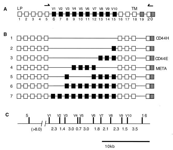
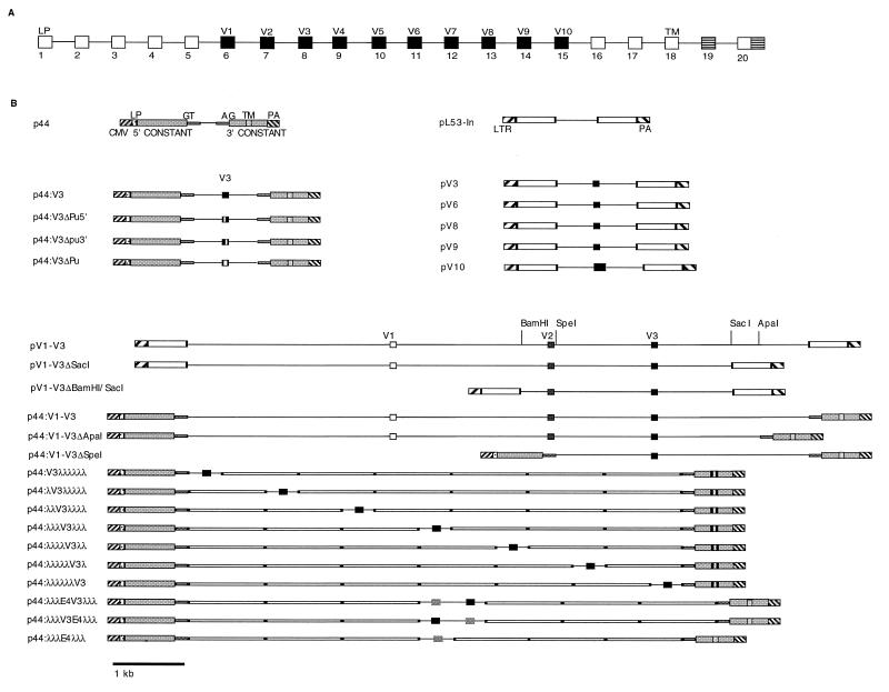
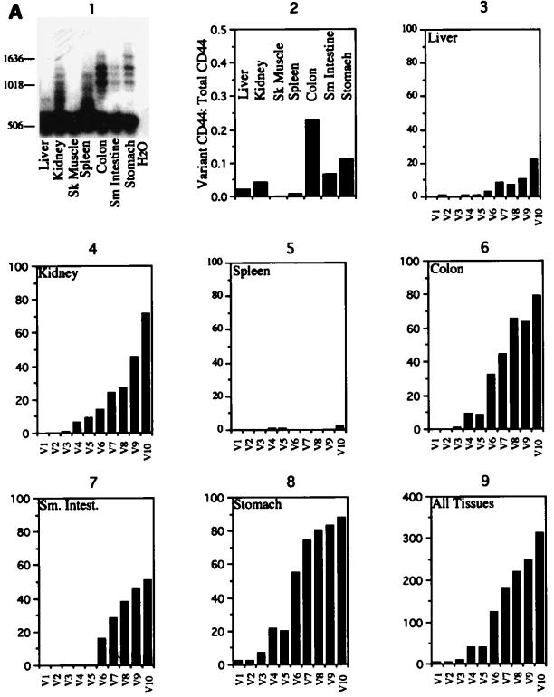
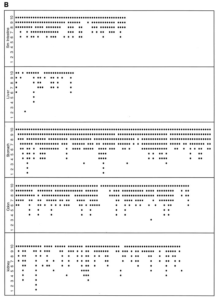
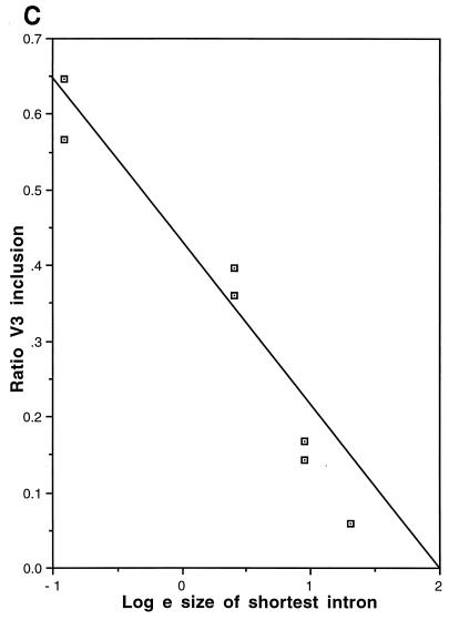
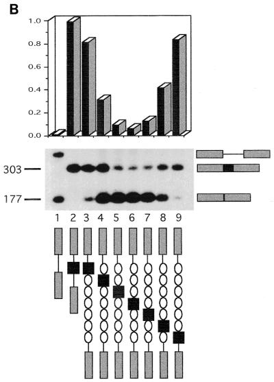
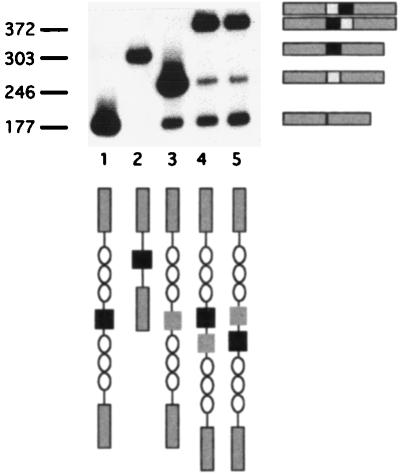
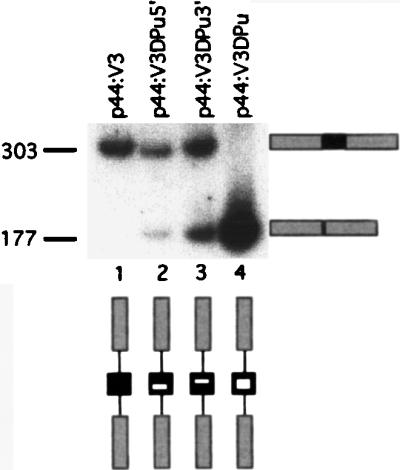
Although the splicing of transcripts from most eukaryotic genes occurs in a constitutive fashion, some genes can undergo a process of alternative splicing. This is a genetically economical process which allows a single gene to give rise to several protein isoforms by the inclusion or exclusion of sequences into or from the mature mRNA. CD44 provides a unique example; more than 1,000 possible isoforms can be produced by the inclusion or exclusion of a central tandem array of 10 alternatively spliced exons. Certain alternatively spliced exons have been ascribed specific functions; however, independent regulation of the inclusion or skipping of each of these exons would clearly demand an extremely complex regulatory network. Such a network would involve the interaction of many exon-specific trans-acting factors with the pre-mRNA. Therefore, to assess whether the exons are indeed independently regulated, we have examined the alternative exon content of a large number of individual CD44 cDNA isoforms. This analysis shows that the downstream alternatively spliced exons are favored over those lying upstream and that alternative exons are often included in blocks rather than singly. Using a novel in vivo alternative splicing assay, we show that intron length has a major influence upon the alternative splicing of CD44. We propose a kinetic model in which short introns may overcome the poor recognition of alternatively spliced exons. These observations suggest that for CD44, intron length has been exploited in the evolution of the genomic structure to enable tissue-specific patterns of splicing to be maintained.


Figures

FIG. 1
FIG. 1
Alternative splicing of CD44. (A) Map of the CD44 gene. Exon 1 encodes the leader peptide (LP), and exon 18 encodes the transmembrane domain (TM). Exons 6 to 15 correspond to the alternatively spliced exons (V1 to V10). Exon 19 encodes a short cytoplasmic domain (32). (B) Examples of some previously observed isoforms of CD44: 1, basic, or hematopoietic, variant CD44H; 2, V10 only; 3, epithelial variant (35); 4, META-1 variant (13). (C) Scale map of the genomic locus encompassing the alternatively spliced exons. Intron sizes are given in kilobases. The exon 5-V1 intron size has not been directly measured but is at least 8 kb.
FIG. 2
FIG. 2
Plasmid constructs. (A) Exon map of the CD44 gene. (B) Diagrammatic representation of the constructs used in this work. pV3, pV6, pV8, pV9, and pV10 were constructed in pL53-In. For p44 and its derivatives, shaded areas represent the 5′ and 3′ constant regions of human CD44, consisting of exons 1 to 5 and exons 16 to 18 or 20, respectively. CMV, cytomegalovirus promoter; LTR, Rous sarcoma virus long terminal repeat promoter; LP, leader peptide; GT, start of intron following exon 5; AG, end of intron prior to exon 15; TM, transmembrane domain; PA, polyadenylation signal.
FIG. 3
FIG. 3
Murine CD44 splicing patterns. (A) CD44 was amplified from normal mouse tissue cDNAs and, following Southern transfer, probed with an oligonucleotide which detects all CD44 isoforms (panel 1). A total of 1,000 to 2,000 CD44-containing clones from each tissue were analyzed. The majority of these clones were CD44H, containing no alternative exons (panel 2). The exon compositions of 328 alternative exon-containing variants were determined (panels 3 to 8) (liver [n = 28], kidney [n = 77], spleen [n = 4], colon [n = 80], small (Sm) intestine (Intest) [n = 51], and stomach [n = 88]). No alternatively spliced isoforms were observed in skeletal (Sk) muscle. (B) Schematic of the alternative exon compositions of alternatively spliced cDNA clones included in this study. The numbers at the left represent alternative exons V1 to V10. Exons scored as present are indicated by black dots.
FIG. 3
FIG. 3
Murine CD44 splicing patterns. (A) CD44 was amplified from normal mouse tissue cDNAs and, following Southern transfer, probed with an oligonucleotide which detects all CD44 isoforms (panel 1). A total of 1,000 to 2,000 CD44-containing clones from each tissue were analyzed. The majority of these clones were CD44H, containing no alternative exons (panel 2). The exon compositions of 328 alternative exon-containing variants were determined (panels 3 to 8) (liver [n = 28], kidney [n = 77], spleen [n = 4], colon [n = 80], small (Sm) intestine (Intest) [n = 51], and stomach [n = 88]). No alternatively spliced isoforms were observed in skeletal (Sk) muscle. (B) Schematic of the alternative exon compositions of alternatively spliced cDNA clones included in this study. The numbers at the left represent alternative exons V1 to V10. Exons scored as present are indicated by black dots.
FIG. 4
FIG. 4
Shortening of introns activates the splicing of V3. RT-PCR analysis of splicing from the heterologous reporter pL53-In is shown. Lane 1, no insert; lane 2, 9-kb genomic fragment of CD44 with exons V1 to V3; lanes 3 to 5, shortening of the introns surrounding V3. Fragment sizes are indicated in base pairs. The fragment migrating at 550 bp in lane 3 was shown by Southern analysis to contain either exon V3 or exon V2.
FIG. 5
FIG. 5
In vivo splicing with a CD44 reporter. (A) Scatter plots from FACS analyses of V3 splicing from p44, which has no insert; p3-10cDNA, a CD44 cDNA control containing all exons from V3 to V10; p44:V3, V3 surrounded by short, 400-bp introns; and p44:V1-V3, containing a 9-kbp genome DNA fragment encompassing exons V1 to V3. Total CD44 is shown on the y axis; V3 is shown on the x axis. Cells positive for both are in the upper right quadrant; V3-negative, CD44-positive cells (V3 skipping) are in the upper left quadrant; and cells negative for both (untransfected) are in the lower left quadrant. (B) Histogram illustrating the data from panel A and the results obtained with the 9-kb p44V1-V3 genomic insert and two restriction fragments shortening the introns surrounding V3. Bars represent V3-positive cells expressed as a ratio to total CD44-positive cells.
FIG. 6
FIG. 6
Alternative exons flanked by short introns are spliced constitutively. RT-PCR analysis of splicing from several single-exon reporter constructs surrounded by short, 400-bp introns in pL53-In is shown. Fragment sizes are indicated in base pairs.
FIG. 7
FIG. 7
Effect of intron size on alternative splicing of V3. (A) FACS analysis of splicing is shown. Black box, V3; gray boxes, donor and acceptor sequences; open circles, 1-kb lambda DNA repeats. Column designated cDNA, control cDNA (V3 to V10); column 1, empty vector; column 2, p44:V3 (V3 with short, 400-bp introns); columns 3 to 9, V3 moved sequentially across a 6-kbp lambda stuffer segment. (B) RT-PCR analysis of the same transfectants is shown. The ratio of V3 to total CD44, as measured by laser densitometry, is illustrated graphically. Fragment sizes are indicated in base pairs. (C) V3 inclusion is exponentially related to intron length.
FIG. 7
FIG. 7
Effect of intron size on alternative splicing of V3. (A) FACS analysis of splicing is shown. Black box, V3; gray boxes, donor and acceptor sequences; open circles, 1-kb lambda DNA repeats. Column designated cDNA, control cDNA (V3 to V10); column 1, empty vector; column 2, p44:V3 (V3 with short, 400-bp introns); columns 3 to 9, V3 moved sequentially across a 6-kbp lambda stuffer segment. (B) RT-PCR analysis of the same transfectants is shown. The ratio of V3 to total CD44, as measured by laser densitometry, is illustrated graphically. Fragment sizes are indicated in base pairs. (C) V3 inclusion is exponentially related to intron length.
FIG. 8
FIG. 8
V3 splicing is rescued by proximity to a “strong” exon. Exon 4 of CD44 (light gray boxes) was cloned on either side of V3 (dark gray boxes) in the p44:λλλV3λλλ construct, and data were obtained by RT-PCR. Fragment sizes are indicated in base pairs.
FIG. 9
FIG. 9
Mutation of a putative ERE affects V3 splicing. RT-PCR analysis of splicing from constructs in which the purine-rich stretch of V3 was replaced by pyrimidines is shown. Lane 1, unmutated pV3; lanes 2 and 3, mutation of 5′ and 3′ halves, respectively; lane 4, double mutant. Fragment sizes are indicated in base pairs.

Similar articles

Cited by

References

    1. Arch R, Wirth K, Hofmann M, Ponta H, Matzku S, Herrlich P, Zoller M. Participation in normal immune responses of a metastasis-inducing splice variant of CD44. Science. 1992;257:682–685. - PubMed
    1. Aruffo A, Stamenkovic I, Melnick M, Underhill C B, Seed B. CD44 is the principal cell surface receptor for hyaluronate. Cell. 1990;61:1303–1313. - PubMed
    1. Auch D, Reth M. Exon trap cloning: using PCR to rapidly detect and clone exons from genomic DNA fragments. Nucleic Acids Res. 1990;18:6743–6744. - PMC - PubMed
    1. Bartolazzi A, Jackson D, Bennett K, Aruffo A, Dickinson R, Shields J, Whittle N, Stamenkovic I. Regulation of growth and dissemination of a human lymphoma by CD44 splice variants. J Cell Sci. 1995;108:1723–1733. - PubMed
    1. Bennett K L, Jackson D G, Simon J C, Tanczos E, Peach R, Modrell B, Stamenkovic I, Plowman G, Aruffo A. CD44 isoforms containing exon V3 are responsible for the presentation of heparin-binding growth factor. J Cell Biol. 1995;128:687–698. - PMC - PubMed

Publication types

LinkOut - more resources