. 1998 Oct;18(10):5992-6000.
doi: 10.1128/MCB.18.10.5992.

The locus control region is necessary for gene expression in the human beta-globin locus but not the maintenance of an open chromatin structure in erythroid cells

Affiliations

The locus control region is necessary for gene expression in the human beta-globin locus but not the maintenance of an open chromatin structure in erythroid cells

A Reik et al. Mol Cell Biol. 1998 Oct.

Abstract

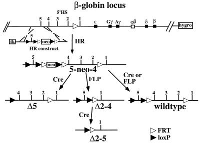
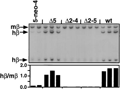
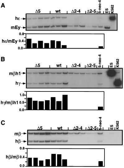
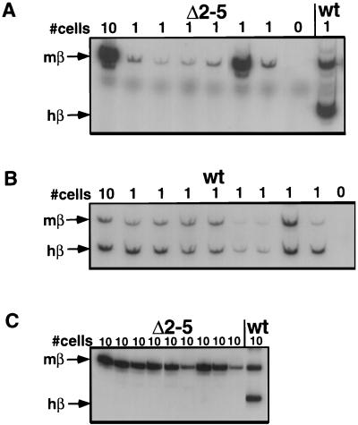
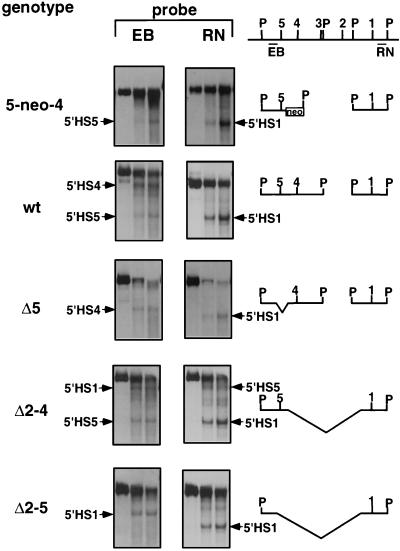
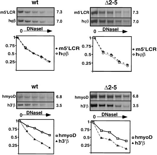
Studies in many systems have led to the model that the human beta-globin locus control region (LCR) regulates the transcription, chromatin structure, and replication properties of the beta-globin locus. However the precise mechanisms of this regulation are unknown. We have developed strategies to use homologous recombination in a tissue culture system to examine how the LCR regulates the locus in its natural chromosomal environment. Our results show that when the functional components of the LCR, as defined by transfection and transgenic studies, are deleted from the endogenous beta-globin locus in an erythroid background, transcription of all beta-globin genes is abolished in every cell. However, formation of the remaining hypersensitive site(s) of the LCR and the presence of a DNase I-sensitive structure of the beta-globin locus are not affected by the deletion. In contrast, deletion of 5'HS5 of the LCR, which has been suggested to serve as an insulator, has only a minor effect on beta-globin transcription and does not influence the chromatin structure of the locus. These results show that the LCR as currently defined is not necessary to keep the locus in an "open" conformation in erythroid cells and that even in an erythroid environment an open locus is not sufficient to permit transcription of the beta-like globin genes.


Figures

FIG. 1
FIG. 1
Homologous recombination in DT40 cells on a modified human chromosome 11 with the human β-globin locus. The genes of the β-globin locus and the upstream HSs are indicated, as well as the modification introduced into the locus in an earlier study (13) (see the text). Homologous recombination creates the 5-neo-4 mutation. After transfer of this chromosome into MEL cells, transient expression of the respective site-specific recombinases creates the indicated mutations. Abbreviations: neo, neomycin resistance gene; hygro, hygromycin B resistance gene; tk, thymidine kinase gene for negative selection. FLP recombinase recognition sites are depicted as open triangles; Cre recombinase recognition sites are depicted as solid triangles.
FIG. 2
FIG. 2
RT-PCR analysis of induced β-globin transcription in the LCR mutations in MEL cells. RT-PCR was performed with a primer pair which coamplifies the human and murine adult β-globin mRNA followed by restriction enzyme digestion specific for the human product and gel electrophoresis. Individual lines with the indicated genotype were analyzed (the second 5-neo-4 line shown is a subclone of the 5-neo-4 line used to create the LCR mutations that was transfected with FLP but did not carry out a recombination event). The ratio of human to mouse signal is shown below the autoradiograph. Note that the weak bands that appear above the main β-globin bands are proportional in their intensities to the correct bands. wt, wild type.
FIG. 3
FIG. 3
RT-PCR analysis of induced β-globin transcription in GM979 cells. RT-PCR was performed as in Fig. 2 with primer pairs which coamplify the murine and human β-globin mRNAs followed by a species-specific restriction digest. (A) Coamplification of human ɛ-globin and mouse Ey. (B) Human γ-globin and mouse βh1 expression. (C) Adult human and murine β-globin expression. The ratio of human to murine signal after quantitation is shown below each lane. ES and K562 are controls for mouse- and human-specific signals and the restriction enzyme digestion. wt, wild type.
FIG. 4
FIG. 4
Single-cell PCR analysis of LCR mutations in MEL cells. Sorted induced MEL cells in the numbers indicated at the top were analyzed in an RT-PCR assay that was able to detect β-globin transcription from single cells. Adult murine and human β-globin mRNAs are amplified and distinguished by restriction enzyme digestion as indicated on the left. (A) Analysis of single MEL cells carrying the Δ2–5 deletion. (B) Analysis of single MEL cells with wild-type (wt) human chromosome 11. (C) Analysis of 10-cell pools carrying the Δ2–5 deletion.
FIG. 5
FIG. 5
DNase I HS analysis of LCR mutations in MEL cells. Cells were digested with increasing concentrations of DNase I, and DNA was cut with PvuII and analyzed by hybridization against probes 5′ of 5′HS5 (left, EB) and 3′ of 5′HS1 (right, RN). The HSs detected in the different lines are indicated. Note that because of the deletions, the size of the PvuII band and the locations of the HSs differ between the lines. The fragments which can be observed in the different mutations with the two probes are indicated to the right, with P denoting PvuII sites. 5′HS3 is located so close to a PvuII site that it cannot be distinguished from the PvuII band by this probing strategy. The 5-neo-4 insertion leads to the insertion of a PvuII site between 5′HS5 and 5′HS4. All data shown are from one filter that had been hybridized sequentially to the EB and the RN probes. wt, wild type.
FIG. 6
FIG. 6
General DNase I sensitivity of the human β-globin locus. Nuclei were digested with increasing amounts of DNase I, and DNA was purified, cut with EcoRI, and analyzed by Southern blotting. Hybridization against the probes indicated on the left of the panel was performed. (The sizes of the resulting bands in kilobases are given on the right.) Quantitation, with the undigested band set at 1, is shown below. Probes: ϕβ and 3′β, in the human ϕβ-globin pseudogene and 3′ of the human β-globin gene; m5′LCR, sensitive region upstream of the murine β-globin LCR; hmyoD, the human myoD gene region on chromosome 11 as a resistant control.

References

    1. Aladjem M I, Groudine M, Brody L L, Dieken E S, Fournier R E K, Wahl G M, Epner E M. Participation of the human β-globin locus control region in initiation of DNA replication. Science. 1995;270:815–819. - PubMed
    1. Amrolia P J, Ramamurthy L, Saluja D, Tanese N, Jane S M, Cunningham J M. The activation domain of the enhancer binding protein p45NF-E2 interacts with TAFII130 and mediates long-range activation of the α- and β-globin gene loci in an erythroid cell line. Proc Natl Acad Sci USA. 1997;94:10051–10056. - PMC - PubMed
    1. Ashe H L, Monks J, Wijgerde M, Fraser P, Proudfoot N J. Intergenic transcription and transinduction of the human β-globin locus. Genes Dev. 1997;11:2494–2509. - PMC - PubMed
    1. Bender, M. Unpublished data.
    1. Bender, M., A. Reik, J. Close, A. Telling, E. Epner, S. Fiering, R. Hardison, and M. Groudine. Description and targeted deletion of 5′HS 5 and 6 of the mouse β-globin locus control region. Blood, in press. - PubMed

Publication types

LinkOut - more resources