. 1998 Oct;18(10):6075-82.
doi: 10.1128/MCB.18.10.6075.

Biased suppression of hematopoiesis and multiple developmental defects in chimeric mice containing Shp-2 mutant cells

Affiliations

Biased suppression of hematopoiesis and multiple developmental defects in chimeric mice containing Shp-2 mutant cells

C K Qu et al. Mol Cell Biol. 1998 Oct.

Abstract

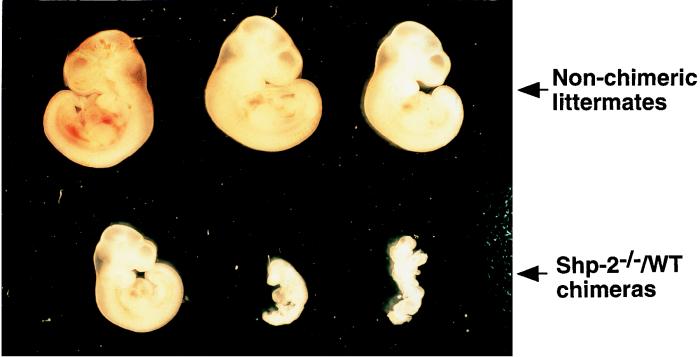
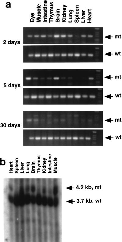
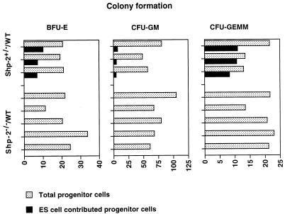
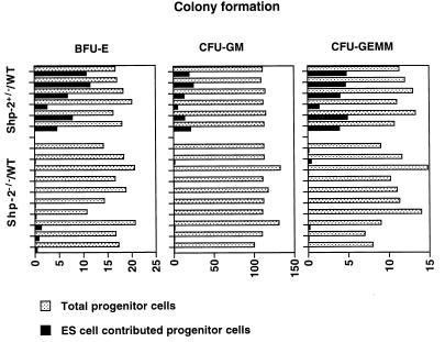
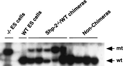
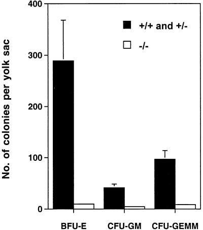
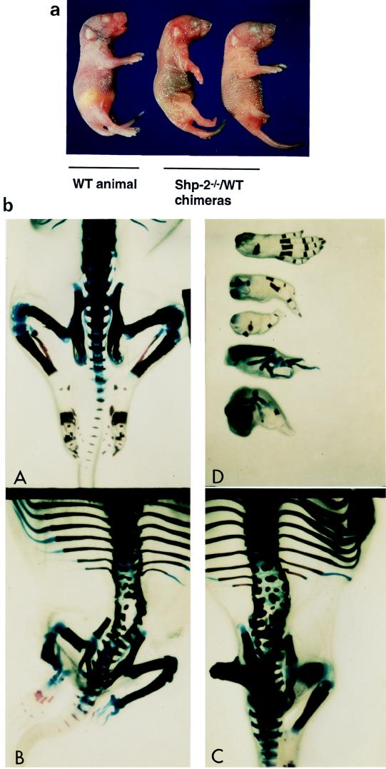
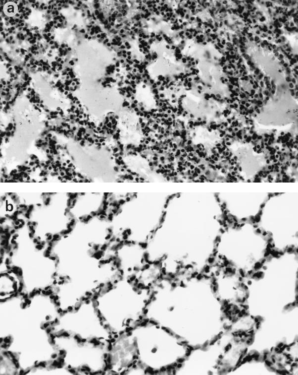
Shp-2 is a cytoplasmic tyrosine phosphatase that contains two Src homology 2 (SH2) domains at the N terminus. Biochemical data suggests that Shp-2 acts downstream of a variety of receptor and cytoplasmic tyrosine kinases. A targeted deletion mutation in the N-terminal SH2 (SH2-N) domain results in embryonic lethality of homozygous mutant mice at midgestation. In vitro embryonic stem (ES) cell differentiation assays suggest that Shp-2 might play an important role in hematopoiesis. By aggregating homozygous mutant (Shp-2(-/-)) ES cells and wild-type (WT) embryos, we created Shp-2(-/-)-WT chimeric animals. We report here an essential role of Shp-2 in the control of blood cell development. Despite the widespread contribution of mutant cells to various tissues, no Shp-2(-/-) progenitors for erythroid or myeloid cells were detected in the fetal liver and bone marrow of chimeric animals by using the in vitro CFU assay. Furthermore, hematopoiesis was defective in Shp-2(-/-) yolk sacs. In addition, the Shp-2 mutation caused multiple developmental defects in chimeric mice, characterized by short hind legs, aberrant limb features, split lumbar vertebrae, abnormal rib patterning, and pathological changes in the lungs, intestines, and skin. These results demonstrate a functional involvement of Shp-2 in the differentiation of multiple tissue-specific cells and in body organization. More importantly, the requirement for Shp-2 is more stringent in hematopoiesis than in other systems.


Figures

FIG. 1
FIG. 1
Heavily chimeric Shp-2−/−–WT embryos were embryo lethal at midgestation. Pregnant foster females at 10.5 d.p.c. were sacrificed, and embryos were dissected from maternal tissues and photographed under a dissecting microscope, followed by genomic DNA extraction and PCR analysis. Chimerism was determined by PCR detection of the Shp-2 mutant allele in embryonic cells.
FIG. 2
FIG. 2
Contribution of Shp-2−/− cells to various tissues in chimeric mice. (a) Three chimeric mice that survived for 2, 5, and 30 days were dissected. Genomic DNA was extracted from the eyes, muscle, intestine, thymus, brain, kidney, lung, spleen, liver, and heart. Shp-2 mutant (mt) and WT (wt) alleles were detected by PCR with specific pairs of primers (29). The density of DNA bands reflects the relative contributions of Shp-2−/− ES cells versus those of the parental embryo. (b) Tissues from a chimeric pup were dissected, and isolated genomic DNA was subjected to Southern blot analysis with a [α-32P]dCTP-labelled specific probe (29). The relative contributions from mutant cells and the parental embryo were determined by comparing the densities of a 4.2-kb mutant DNA band and a 3.7-kb WT DNA band.
FIG. 3
FIG. 3
Detection of hematopoietic progenitors in the bone marrow of Shp-2−/−–WT chimeras. Bone marrow cells of two femurs from each of five Shp-2−/−–WT and three Shp-2+/−–WT chimeras were flushed out and disaggregated into single-cell suspensions. Nucleated cells (5.0 × 10⁴/ml) were seeded into semisolid methylcellulose CFU assay culture systems with and without G418 at a concentration of 1.25 mg/ml, as described in the text. After 6 to 7 days of incubation, colonies of BFU-E, CFU-GM, and CFU-GEMM cells grown in cultures without G418 were scored as total progenitor cells, and colonies grown in G418-containing medium were enumerated as mutant ES cell-derived progenitor cells.
FIG. 4
FIG. 4
Definitive hematopoietic cell development in chimeric fetal livers. Shp-2−/−–WT and Shp-2+/−–WT chimeric embryos were dissected at 14.5 d.p.c. Individual fetal livers were isolated in α-MEM plus 15% FCS and disaggregated mechanically into single-cell suspensions. Nucleated cells (2.5 × 10⁴/ml) were plated for CFU assays as described in the legend to Fig. 3. After 6 to 7 days of culture, total and ES cell-contributed BFU-E, CFU-GM, and CFU-GEMM progenitor cells were scored for each fetal liver.
FIG. 5
FIG. 5
Contribution of Shp-2−/− cells in whole embryos. Chimeric and nonchimeric embryos were dissected at 14.5 d.p.c. and subjected to genomic DNA extraction. Southern blot analysis was performed for determination of the relative contribution of Shp-2−/− cells versus that of the parental embryo, as described in the legend to Fig. 2b. Densitometry of the WT (wt) and mutant (mt) bands showed that the percent contribution from ES cells for the five chimeras, from left to right, was 31.1, 35.1, 44.8, 63.0, and 41.4.
FIG. 6
FIG. 6
Defective hematopoiesis in Shp-2−/− yolk sacs. Yolk sacs at 8.5 to 9.0 d.p.c. from Shp-2+/− × Shp-2+/− crosses were carefully dissected free of maternal tissues under sterile conditions in α-MEM plus 15% FCS and were disaggregated by passing them through a 22-gauge needle. After being washed once in α-MEM, whole yolk sac cells were plated for the CFU assay. Error bars indicate standard deviations.
FIG. 7
FIG. 7
Abnormalities of terminal and skeletal structures in Shp-2−/−–WT chimeras. (a) Examples from one nonchimeric animal and two chimeric animals are shown. Chimeras were recognized by agouti eye color at birth. The abnormality in hind-leg development was obvious. (b) Newborn Shp-2−/−–WT chimeric pups and nonchimeric controls were eviscerated and fixed in 100% ethanol and acetone. Skeletal specimens were prepared as described in the text. Structural changes were examined and photographed under a dissecting microscope. (c) WT animal; (A and D) typical representatives of Shp-2−/−–WT chimeras; (B) WT mouse hind foot with five fingers and four different abnormal feet from Shp-2−/−–WT chimeric mice.
FIG. 8
FIG. 8
Histopathological changes in the lungs of Shp-2−/−–WT chimeric mice. (a) Mutant lung section, with hypercellular thickened alveolar walls; (b) lung of normal newborn, with thin alveolar walls and well-developed alveolar spaces. Magnification, ×100. Hematoxylin and eosin staining was used.

References

    1. Bash R O, Hall S, Timmons C F, Crist W M, Amylon M, Smith R G, Baer R. Does activation of the TAL1 gene occur in a majority of patients with T-cell acute lymphoblastic leukemia? A pediatric oncology group study. Blood. 1995;86:666–676. - PubMed
    1. Cooper, S., and H. E. Broxmeyer. 1996. Measurement of interleukin-3 and other hematopoietic growth factors, such as GM-CSF, G-CSF, M-CSF, erythropoietin and the other potent co-stimulating cytokines steel factor and Flt-3 ligand. Curr. Protocols Immunol. 18(Suppl.):6.4.1-6.4.12. - PubMed
    1. Deng C X, Wynshaw-Boris A, Shen M M, Daugherty C, Ornitz D M, Leder P. Murine FGFR-1 is required for early postimplantation growth and axial organization. Genes Dev. 1994;8:3045–3057. - PubMed
    1. de Vries C, Escobedo J A, Ueno H, Houck K, Ferrara N, Williams L T. The fms-like tyrosine kinase, a receptor for vascular endothelial growth factor. Science. 1992;255:989–991. - PubMed
    1. Feng G S, Hui C C, Pawson T. SH2-containing phosphotyrosine phosphatase as a target of protein-tyrosine kinases. Science. 1993;259:1607–1611. - PubMed

Publication types

Substances

LinkOut - more resources