Comparative Study
. 1998 Oct 1;18(19):7674-86.
doi: 10.1523/JNEUROSCI.18-19-07674.1998.

The cellular and subcellular localization of huntingtin-associated protein 1 (HAP1): comparison with huntingtin in rat and human

Affiliations
Comparative Study

The cellular and subcellular localization of huntingtin-associated protein 1 (HAP1): comparison with huntingtin in rat and human

C A Gutekunst et al. J Neurosci. .

Abstract

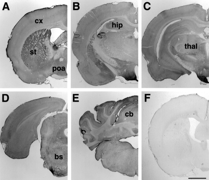
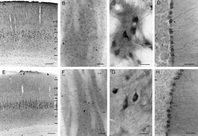
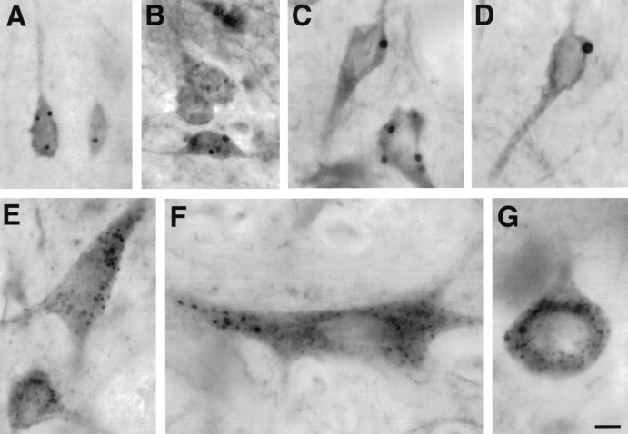
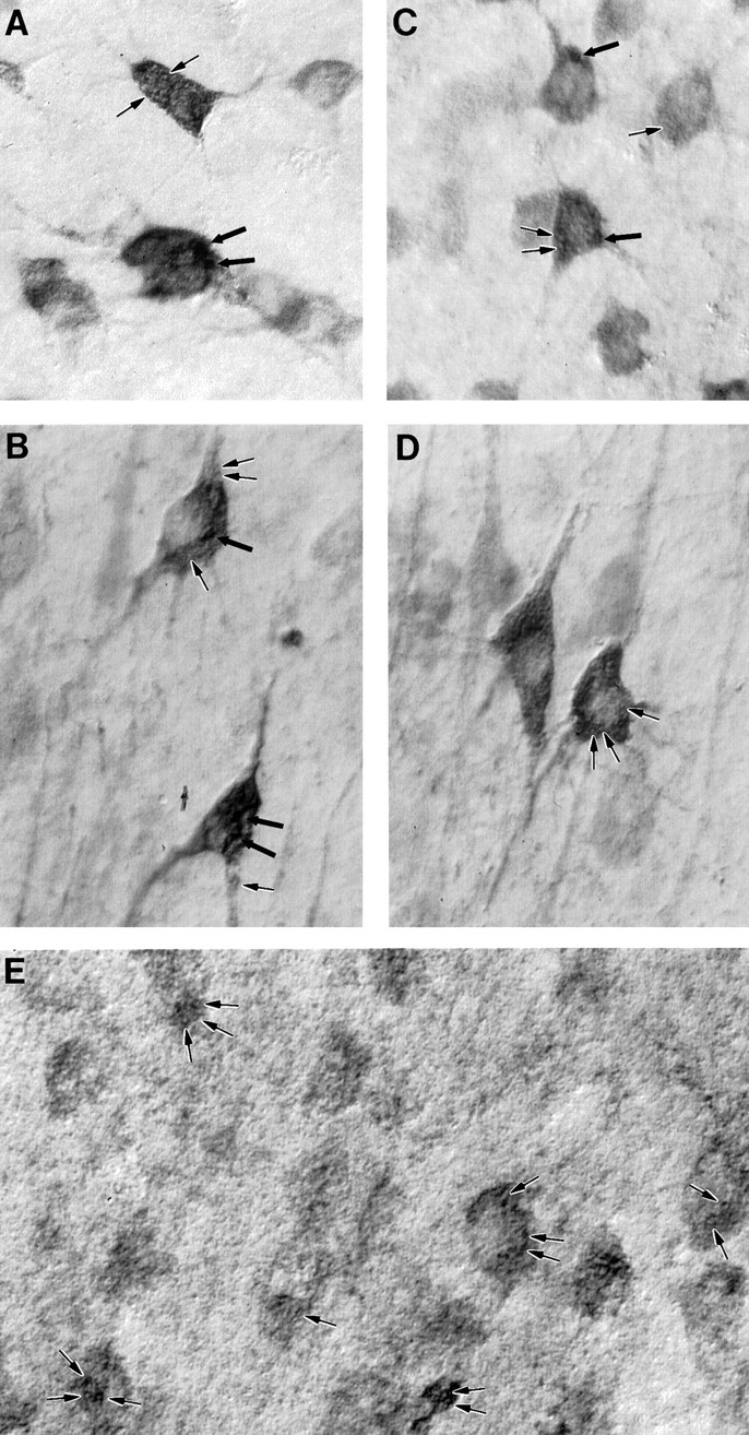
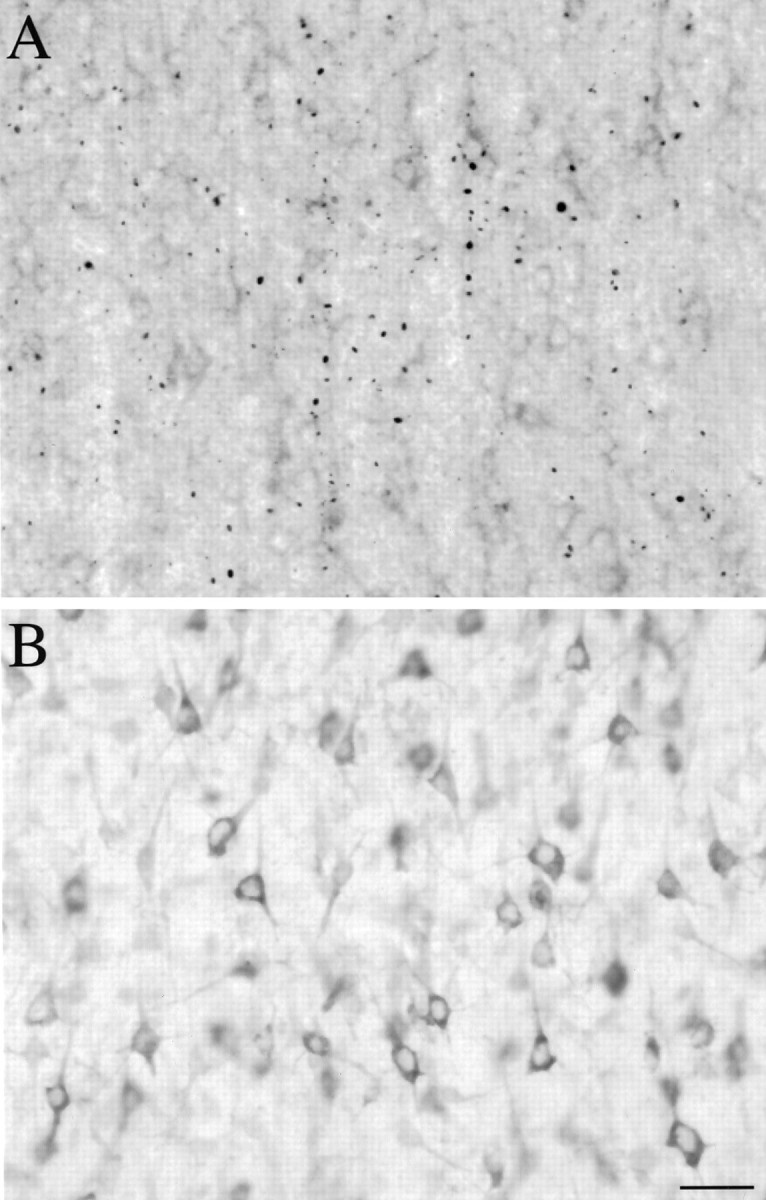
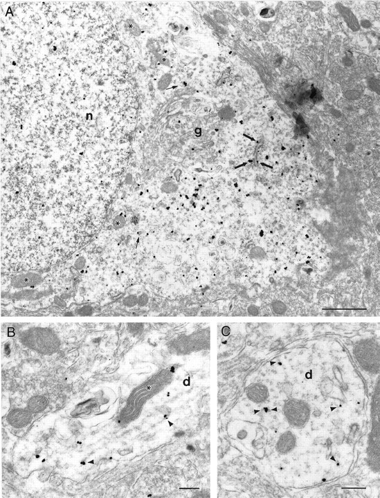
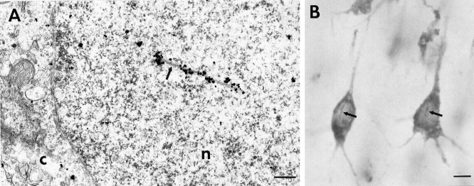
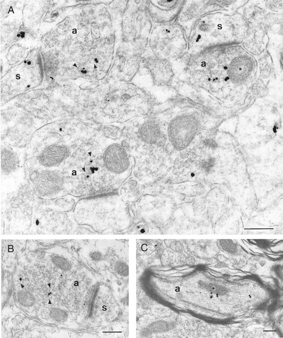
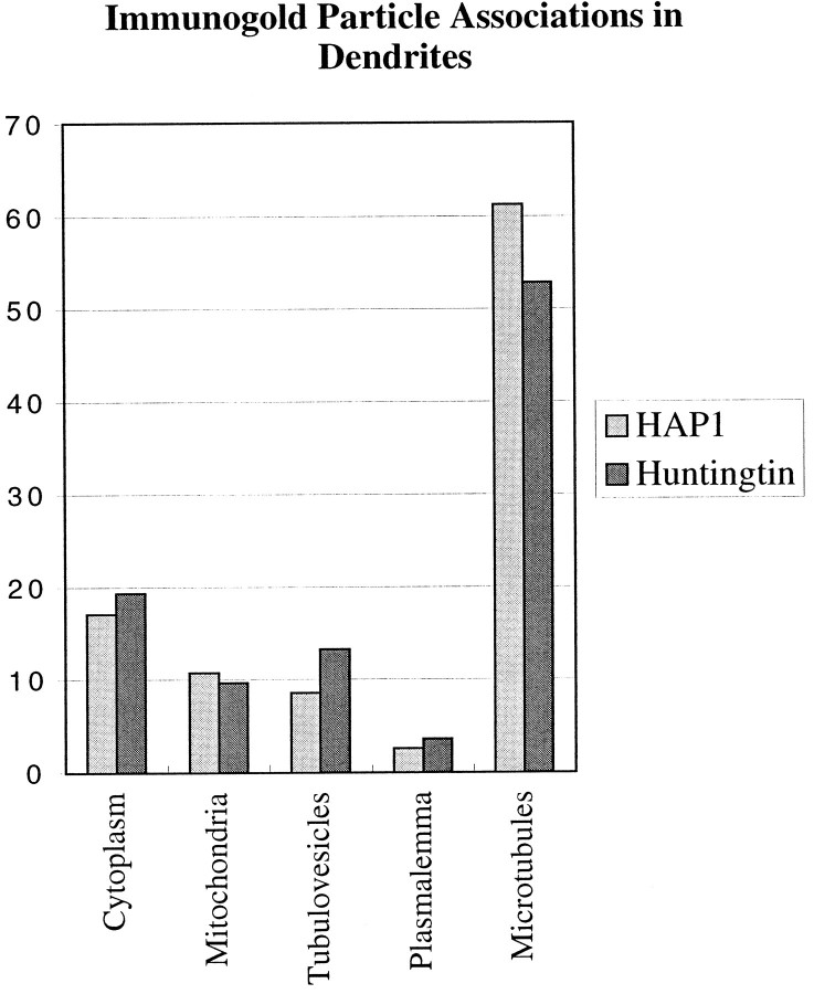
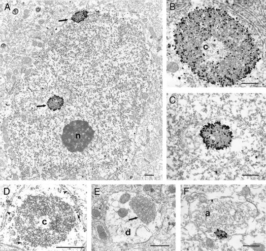
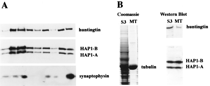
The cellular and subcellular distribution of HAP1 was examined in rat brain by light and electron microscopic immunocytochemistry and subcellular fractionation. HAP1 localization was also determined in human postmortem tissue from control and Huntington's disease (HD) cases by light microscopic immunocytochemistry. At the cellular level, the heterogeneity of HAP1 expression was similar to that of huntingtin; however, HAP1 immunoreactivity was more widespread. The subcellular distribution of HAP1 was examined using immunogold electron microscopy. Like huntingtin, HAP1 is a cytoplasmic protein that associates with microtubules and many types of membranous organelles, including mitochondria, endoplasmic reticulum, tubulovesicles, endosomal and lysosomal organelles, and synaptic vesicles. A quantitative comparison of the organelle associations of HAP1 and huntingtin showed them to be almost identical. Within HAP1-immunoreactive neurons in rat and human brain, populations of large and small immunoreactive puncta were visible by light microscopy. The large puncta, which were especially evident in the ventral forebrain, were intensely HAP1 immunoreactive. Electron microscopic analysis revealed them to be a type of nucleolus-like body, which has been named a stigmoid body, that may play a role in protein synthesis. The small puncta, less intensely labeled, were primarily mitochondria. These results indicate that the localization of HAP1 and huntingtin is more similar than previously appreciated and provide further evidence that HAP1 and huntingtin have localizations consistent with roles in intracellular transport. Our data also suggest, however, that HAP1 is not present in the abnormal intranuclear and neuritic aggregates containing the N-terminal fragment of mutant huntingtin that are found in HD brains.


Figures

Fig. 1.
Fig. 1.
Regional distribution of HAP1 in the rat brain.A–E, Representative coronal sections from HAP1-immunostained tissue are shown from rostral to caudal.F, The immunolabeling is abolished after preabsorption over excess GST–HAP1 fusion protein. bs, Brainstem;cb, cerebellum; cx, cortex;hip: hippocampus; poa, preoptic area;st, striatum thal, thalamus. Scale bar, 1 mm.
Fig. 2.
Fig. 2.
Similarities in HAP1 and huntingtin distribution in the rat brain. Light micrographs showing HAP1 (A–D) and huntingtin Hp549 (E–H) immunoreactivity are shown. Both HAP1 and huntingtin immunolabeling filled neuronal perikarya and dendrites. Throughout the brain, HAP1 cellular distribution was very similar to that of huntingtin; however HAP1 labeled more neurons than did huntingtin. A, E, In somatosensory cortex, HAP1 (A) and huntingtin (E) are found in pyramidal neurons.B, F, In striatum, both medium-sized neurons and larger neurons (arrows) are labeled with HAP1 (B) and huntingtin (F). C, G, In globus pallidus, neurons and dendrites are well stained by both HAP1 (C) and huntingtin (G).D, H, In the cerebellum, Purkinje cells (arrows) are more intensely stained by both HAP1 (D) and huntingtin (H) than are neurons in the granule cell layer (g). Scale bars: A,E, 200 μm; B, D,F, H, 50 μm; C,G, 25 μm.
Fig. 3.
Fig. 3.
HAP1 labeling of large and small inclusions in the rat brain. Light micrographs showing HAP1 immunolabeling of inclusions in neurons from the cortex (A), striatum (B), preoptic area (C), and the septal region (D) are shown. In globus pallidus (E), brainstem neurons (F), and Purkinje cells (G), HAP1 immunoperoxidase labels organelles that are smaller and more numerous than stigmoid bodies and may primarily be mitochondria. Scale bar, 10 μm.
Fig. 4.
Fig. 4.
HAP1 immunostaining in the human and comparison with huntingtin and mitochondrial membrane markers. Nomarski imaging of HAP1 (A, B), huntingtin (C, D), and mitochondria (E) in human brain is shown. Large and small inclusions (thick and thin arrows, respectively), similar to those visualized in the rat, are visible in these neurons from the putamen (A, C) and cerebral cortex (B, D). The human mitochondrial marker MAB1273 is shown (E) labeling inclusions (arrows) similar in size to the small HAP1- and huntingtin-immunoreactive inclusions.
Fig. 5.
Fig. 5.
HAP1 is not present in N-terminal huntingtin aggregates. A, Light micrograph showing EM48 labeling in cortex from an HD case. Em48 immunostained many aggregates located mostly in the neuropil but also found in neuronal nuclei.B, An adjacent section immunolabeled with HAP1. Immunostaining is localized to neuronal perikarya and proximal processes and is diffuse in the neuropil. There is no labeling of aggregates. Scale bar, 20 μm.
Fig. 6.
Fig. 6.
Subcellular localization of HAP1.A, Electron micrograph showing the cell body of a HAP1-immunolabeled neuron in the septal region. Most immunogold particles were cytoplasmic, whereas a few were also found in the nucleus (n). Most particles were either free in the cytoplasm in regions rich in polysomes or in contact with the outer surface of rough endoplasmic reticulum (thick arrows), mitochondria (asterisks), and tubulovesicular elements (thin arrows). A few rare particles were found in contact with the plasma membrane or within regions rich in Golgi apparatus (g). B, Longitudinal section through a dendrite (d) showing immunogold particles in contact with microtubules (arrowheads) or a mitochondrion (asterisk). C, Cross-section through a dendrite (d) in which immunogold particles can be seen in contact with microtubule cross-sections (arrowheads). Scale bars, 250 nm.
Fig. 7.
Fig. 7.
Association of HAP1 to nuclear rods.A, Electron micrograph showing HAP1 immunogold in the nucleus (n) of a preoptic area neuron. Gold particles are located along an intranuclear rod (arrow). The cytoplasm (C) is separated from the nucleus by the nuclear envelope. B, Light micrograph of a coronal section through normal human cortex showing HAP1 immunostaining. In pyramidal neurons, the DAB reaction product is present in perikarya and proximal dendrites but is also found along intranuclear rods (arrows). Scale bars:A, 300 nm; B, 10 μm.
Fig. 8.
Fig. 8.
Subcellular localization of HAP1.A, B, Electron micrographs showing HAP1 immunogold particles in four axon terminals (a) and in three of their postsynaptic dendritic spines (s). In the axon terminals, immunogold particles are located amid the synaptic vesicles, frequently contacting their cytoplasmic surfaces (arrowheads) but also contacting the outer membrane of mitochondria (asterisk). Immunogold particles in dendritic spines were free in the cytoplasm. In both axon terminals and dendritic spines, immunogold particles were not found in association with synaptic junctional complexes.C, Electron micrograph of a myelinated axon (a) showing a few immunogold particles either amid cytoskeletal elements or in contact with a mitochondrion (asterisk). Scale bars, 250 nm.
Fig. 9.
Fig. 9.
Comparison of the proportional labeling of organelles and membranes by HAP1 and huntingtin immunogold particles. Histograms showing the proportions of immunogold particles observed to be associated with particular subcellular compartments in frontal cortex dendrites are shown. These data indicate that the proportional distribution of HAP1 and huntingtin among the compartments is very similar. Because the frequency and surface areas of each compartment are not comparable, however, these data should not be interpreted as giving any indication of the relative abundance of HAP1 or huntingtin in each.
Fig. 10.
Fig. 10.
Labeled and unlabeled stigmoid bodies in neurons.A–F, Electron micrographs of stigmoid bodies demonstrating their structure, location, and HAP1 immunoreactivity. Stigmoid bodies do not have a limiting membrane and are made up of heterogeneous electron-dense material, often surrounding a large electron-lucent core (c). Much of the dense material appears to be made up of individual rounded electron-dense elements with electron-lucent centers. A, Electron micrograph showing two immunolabeled stigmoid bodies (arrows) in the perikaryon of a neuron in the lateral septum. This perinuclear positioning is very frequent. Immunogold particles are visible both around and within the stigmoid bodies.B, C, Immunogold-labeled stigmoid bodies in neurons from the preoptic area and the lateral septum seen at higher magnification. Label is concentrated on the surface of the stigmoid bodies but also is found deep within the electron-dense material.D, Electron micrograph of unlabeled stigmoid bodies in the perikaryon of a preoptic neuron showing stigmoid ultrastructure unobscured by immunogold particles. E, F, Unlabeled stigmoid (arrow) in a medium caliber dendrite (d) and a labeled stigmoid in an axon terminal (a) demonstrating that they can be found in more distal locations. Scale bars, 500 nm.
Fig. 11.
Fig. 11.
Subcellular distribution of HAP1 and huntingtin.A, Subcellular fractions from rat whole brain were prepared according to the method of Huttner et al. (1983). Western blot analysis was conducted with antibodies for HAP1, huntingtin, and synaptophysin. B, Coomassie blue staining and Western blot analysis of the rat brain microtubule (MT) fraction that was obtained by polymerization of tubulin in the cytosolic fraction (S3) are shown. Western blot analysis was conducted with antibodies to HAP1 and huntingtin.

References

    1. Bao J, Sharp A, Wagster M, Becher M, Schilling G, Ross C, Dawson V, Dawson T. Expansion of polyglutamine repeat in huntingtin leads to abnormal protein interactions involving calmodulin. Proc Natl Acad Sci USA. 1996;93:5037–5042. - PMC - PubMed
    1. Becher MW, Kotzuk JA, Sharp AH, Davies SW, Bates GP, Price DL, Ross CA. Intranuclear neuronal inclusions in Huntington’s disease and dentatorubral and Pallidoluysian atrophy: correlation between the density of inclusions and IT15 CAG triplet repeat length. Neurobiol Dis. 1998;4:387–397. - PubMed
    1. Burke J, Enghild J, Martin M, Jou Y-S, Myers R, Roses A, Vance J, Strittmatter W. Huntingtin and DRPLA proteins selectively interact with the enzyme GAPDH. Nat Med. 1996;2:347–350. - PubMed
    1. Burry R, Vandre D, Hayes D. Silver enhancement of gold antibody probes in pre-embedding electron microscopic immunocytochemistry. J Histochem Cytochem. 1992;40:1849–1856. - PubMed
    1. Davies SW, Turmaine M, Cozens BA, DiFiglia M, Sharp AH, Ross CA, Scherzinger E, Wanker EE, Mangiarini L, Bates GP. Formation of neuronal intranuclear inclusions underlies the neurological dysfunction in mice transgenic for the HD mutation. Cell. 1997;90:537–548. - PubMed

Publication types

MeSH terms