. 1998 Oct 1;18(19):7768-78.
doi: 10.1523/JNEUROSCI.18-19-07768.1998.

Increased neurogenesis in the dentate gyrus after transient global ischemia in gerbils

Affiliations

Increased neurogenesis in the dentate gyrus after transient global ischemia in gerbils

J Liu et al. J Neurosci. .

Abstract

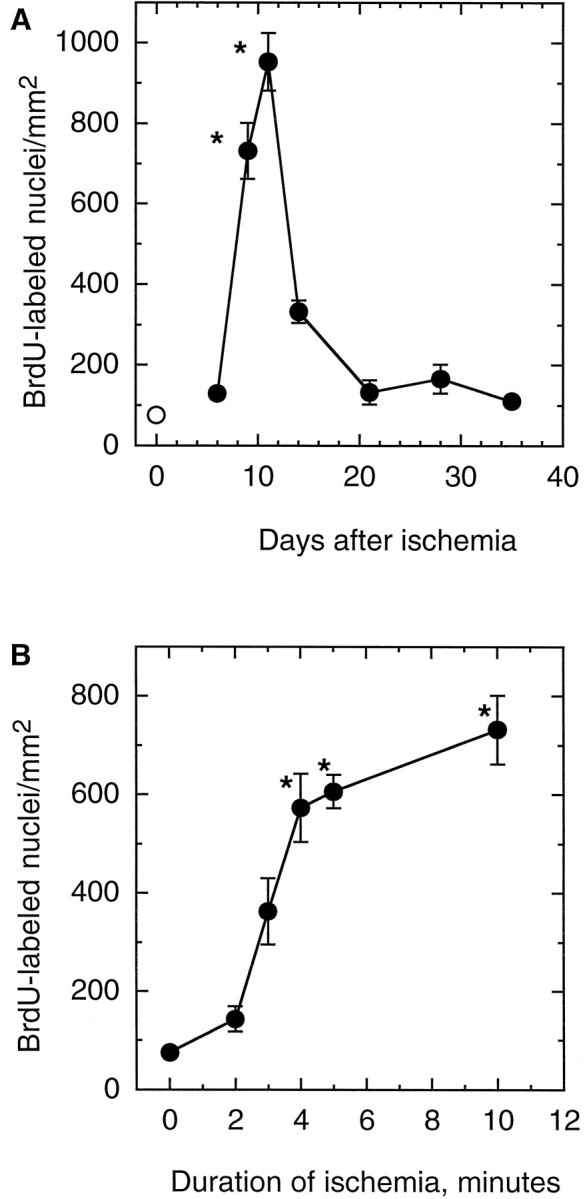
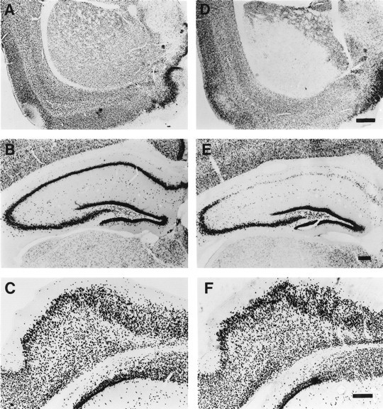
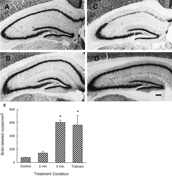
Neurogenesis in the dentate gyrus of adult rodents is regulated by NMDA receptors, adrenal steroids, environmental stimuli, and seizures. To determine whether ischemia affects neurogenesis, newly divided cells in the dentate gyrus were examined after transient global ischemia in adult gerbils. 5-Bromo-2'-deoxyuridine-5'-monophosphate (BrdU) immunohistochemistry demonstrated a 12-fold increase in cell birth in the dentate subgranular zone 1-2 weeks after 10 min bilateral common carotid artery occlusions. Two minutes of ischemia did not significantly increase BrdU incorporation. Confocal microscopy demonstrated that BrdU immunoreactive cells in the granule cell layer colocalized with neuron-specific markers for neuronal nuclear antigen, microtubule-associated protein-2, and calbindin D28k, indicating that the newly divided cells migrated from the subgranular zone into the granule cell layer and matured into neurons. Newborn cells with a neuronal phenotype were first seen 26 d after ischemia, survived for at least 7 months, were located only in the granule cell layer, and comprised approximately 60% of BrdU-labeled cells in the granule cell layer 6 weeks after ischemia. The increased neurogenesis was not attributable to entorhinal cortical lesions, because no cell loss was detected in this region. Ischemic preconditioning for 2 min, which protects CA1 neurons against subsequent ischemic damage, did not prevent increased neurogenesis in the granule cell layer after a subsequent severe ischemic challenge. Thus, ischemia-induced dentate neurogenesis is not attributable to CA1 neuronal loss. Enhanced neurogenesis in the dentate gyrus may be a compensatory adaptive response to ischemia-associated injury and could promote functional recovery after ischemic hippocampal injury.


Figures

Fig. 1.
Fig. 1.
Ischemia increases cell proliferation in the dentate gyrus of adult gerbils. Animals were subjected to 10 min of global ischemia. BrdU was administered 1 d before the animal was killed, and brains were processed for BrdU immunohistochemistry.A, Control untreated gerbil. B–F, Ischemic gerbils killed 8 d (B), 2 weeks (C), 3 weeks (D), 4 weeks (E), or 5 weeks (F) after ischemia. Scale bar, 250 μm.
Fig. 2.
Fig. 2.
Time course of cell proliferation after global ischemia and effect of ischemia duration on proliferation in the SGZ. In both sets of experiments, BrdU was administered 24 hr before the animal was killed. A, The number of BrdU immunoreactive nuclei in the SGZ of animals killed 6 d (n = 12), 9 d (n = 8), 11 d (n = 7), 2 weeks (n = 6), 3 weeks (n = 7), 4 weeks (n = 8), or 5 weeks (n = 6) after 10 min of global ischemia (•). Control animals (n = 10) were untreated (○). B, BrdU-positive nuclei in the SGZ of animals killed 8 d after 2 (n = 8), 3 (n = 6), 4 (n = 6), 5 (n = 6), or 10 (n = 8) min of ischemia. Data are mean ± SEM. Error bars are omitted where they extend beyond the symbols. *p < 0.05 compared with control.
Fig. 3.
Fig. 3.
Neuronal identity of newly divided cells in the dentate gyrus after global ischemia. BrdU was administered twice daily 9–12 d after 10 min of global ischemia. Gerbils were killed 4 weeks later, and brain sections were stained for BrdU immunoreactivity (green) and cell-specific markers (red). A, B, Colocalization of BrdU with calbindin-D28k(CalB) or NeuN is shown by yellow nuclei.C, Colocalization of BrdU with MAP-2 is shown in cells with red cytoplasm surrounding greennuclei. D, No colocalization of GFAP and BrdU was observed within the GCL. Scale bar, 10 μm.
Fig. 4.
Fig. 4.
Distribution and morphology of BrdU-labeled cells after global ischemia. BrdU was given twice daily 9–12 d after ischemia. Animals were then killed 15 (A,F), 26 (B, G), 40 (C, H), or 96 (D,I) d after ischemia. The sham-operated gerbil was killed 40 d (E, J) after surgery. F–J, High-magnification view. Scale bars (inE and F): A–E, 250 μm; F–J, 25 μm.
Fig. 5.
Fig. 5.
Lack of neuronal loss in the entorhinal cortex after 10 min of global ischemia. A–C, Neuronal cells detected in untreated control gerbils by NeuN immunohistochemistry in striatum (A), hippocampus (B), and entorhinal cortex (C). D–F, Animals subjected to 10 min of ischemia and then examined for NeuN immunoreactivity in striatum (D), hippocampus (E), and entorhinal cortex (F) 8 d later. Scale bars: (in D) A, D, 1 mm; (inE) B, E, 200 μm; (inF) C, F, 200 μm.
Fig. 6.
Fig. 6.
Ischemia increases dentate neurogenesis in ischemic-tolerant animals. Animals received 2 (B) or 5 (C) min of global ischemia or 2 min of ischemic preconditioning followed by 5 min of global ischemia (D) compared with untreated control (A). Hippocampal sections were obtained 8 d after the sham operation or the last ischemic insult and analyzed by NeuN immunohistochemistry. Approximately 50% of the preconditioned gerbils showed intact CA1 morphology. Only these animals that lacked CA1 damage were analyzed for dentate neurogenesis. Scale bar, 200 μm.E, Animals were subjected to 2 (n = 8) or 5 (n = 6) min of ischemia. TheTolerant group of animals was subjected to 2 min of ischemic preconditioning, followed by 5 min of ischemia 3 d later (n = 9). BrdU was given 8 d after ischemia, and the animals were killed 1 d later. BrdU was given 24 hr before the control animal was killed (Control) (n = 10). Data shown are mean ± SE. *p < 0.05 compared with control.

References

    1. Altman J, Das G. Postnatal neurogenesis in the guinea-pig. Nature. 1967;214:1098–1101. - PubMed
    1. Bartesaghi R, Gessi T, Migliore M. Input–output relations in the entorhinal–hippocampal–entorhinal loop: entorhinal cortex and dentate gyrus. Hippocampus. 1995;5:440–451. - PubMed
    1. Bayer SA, Yackel JW, Puri PS. Neurons in the rat dentate gyrus granular layer substantially increase during juvenile and adult life. Science. 1982;216:890–892. - PubMed
    1. Bengzon J, Kokaia Z, Elmer E, Nanobashvili A, Kokaia M, Lindvall O. Apoptosis and proliferation of dentate gyrus neurons after single and intermittent limbic seizures. Proc Natl Acad Sci USA. 1997;94:10432–10437. - PMC - PubMed
    1. Bering R, Draguhn A, Diemer NH, Johansen FF. Ischemia changes the co-expression of somatostatin and neuropeptide Y in hippocampal interneurons. Exp Brain Res. 1997;115:423–429. - PubMed

Publication types