. 1998 Oct 1;18(19):7779-99.
doi: 10.1523/JNEUROSCI.18-19-07779.1998.

Regional and cellular patterns of reelin mRNA expression in the forebrain of the developing and adult mouse

Affiliations

Regional and cellular patterns of reelin mRNA expression in the forebrain of the developing and adult mouse

S Alcántara et al. J Neurosci. .

Abstract

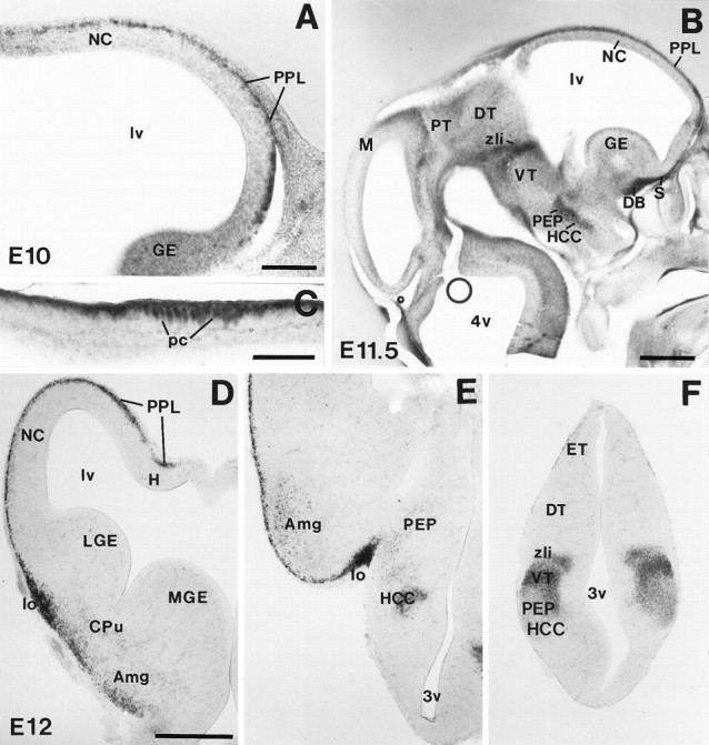
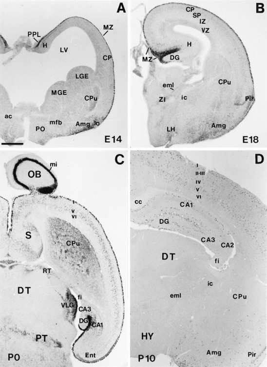
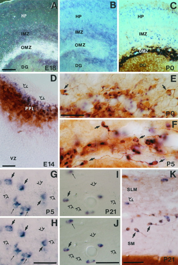
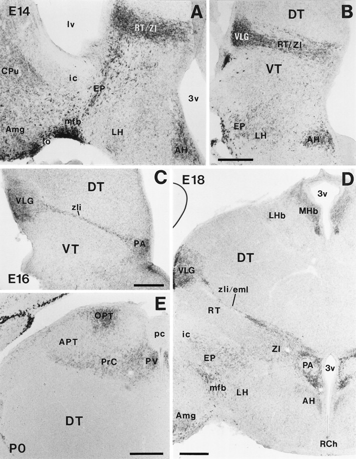
The reelin gene encodes an extracellular protein that is crucial for neuronal migration in laminated brain regions. To gain insights into the functions of Reelin, we performed high-resolution in situ hybridization analyses to determine the pattern of reelin expression in the developing forebrain of the mouse. We also performed double-labeling studies with several markers, including calcium-binding proteins, GAD65/67, and neuropeptides, to characterize the neuronal subsets that express reelin transcripts. reelin expression was detected at embryonic day 10 and later in the forebrain, with a distribution that is consistent with the prosomeric model of forebrain regionalization. In the diencephalon, expression was restricted to transverse and longitudinal domains that delineated boundaries between neuromeres. During embryogenesis, reelin was detected in the cerebral cortex in Cajal-Retzius cells but not in the GABAergic neurons of layer I. At prenatal stages, reelin was also expressed in the olfactory bulb, and striatum and in restricted nuclei in the ventral telencephalon, hypothalamus, thalamus, and pretectum. At postnatal stages, reelin transcripts gradually disappeared from Cajal-Retzius cells, at the same time as they appeared in subsets of GABAergic neurons distributed throughout neocortical and hippocampal layers. In other telencephalic and diencephalic regions, reelin expression decreased steadily during the postnatal period. In the adult, there was prominent expression in the olfactory bulb and cerebral cortex, where it was restricted to subsets of GABAergic interneurons that co-expressed calbindin, calretinin, neuropeptide Y, and somatostatin. This complex pattern of cellular and regional expression is consistent with Reelin having multiple roles in brain development and adult brain function.


Figures

Fig. 1.
Fig. 1.
reelin mRNA expression during early embryonic development. A, Parasagittal section of the telencephalon at E10, showing reelin expression in the outermost preplate layer (PPL) of the neocortex (NC). B, Parasagittal section showing distribution of reelin mRNA in the telencephalon and diencephalon at E11.5. reelin is strongly expressed in thePPL, in the anlages of the septum (S) and diagonal band (DB) complex, in a longitudinal band covering the posterior entopeduncular area (PEP) and the hypothalamic cell cord (HCC), and in three transverse diencephalic domains corresponding to the zona limitans intrathalamica (zli), the border between dorsal thalamus (DT) and pretectum (PT), and in the posterior commissure (pc), at the border between the mesencephalon (M) and the PT. C, High-magnification photomicrograph showing labeled cells at the posterior commissure (pc). D–F, Coronal sections of an E12 embryo showing the distribution ofreelin transcripts at three different rostrocaudal levels (D, rostral; F, caudal). Note the prominent reelin expression associated to the lateral olfactory tract (lo in D, E) and the labeled cells in the presumptive caudate-putamen (CPu), amygdala (Amg), and ventral thalamus (VT). DT, Dorsal thalamus;ET, epithalamus; GE, ganglionic eminence;H, hippocampus; lv, lateral ventricle;LGE, lateral ganglionic eminence; MGE,medial ganglionic eminence; 3v, third ventricle;4v, fourth ventricle. Scale bars: A, C, 150 μm; B, D–F, 500 μm.
Fig. 2.
Fig. 2.
Low-power views illustrating the main features of reelin mRNA distribution from E14 to early postnatal stages. A, Coronal section showing strongreelin expression in the preplate (PPL), marginal zone (MZ), and amygdala (Amg) at E14. B, Coronal section at E18 showing highreelin labeling in the MZ. Very weak staining can be detected at this stage in the cortical plate (CP). C, Horizontal section at P0; the olfactory bulb (OB), cortical layer I (I), hippocampal MZ, and the ventral lateral geniculate nucleus (VLG) show the higher expression levels. High labeling is also found in the caudate-putamen (CPu), in some septal (S) divisions, in the pretectum (PT) at the level of the posterior commissure, and in different cortical and hippocampal layers. D,Coronal section showing the distribution of reelintranscripts at P10. Labeled cells are seen throughout the cortical layers I–VI, hippocampus (H), and Amg. ac, Anterior commissure; CA1, CA2, CA3, hippocampal subfields;cc, corpus callosum; DG, dentate gyrus;DT, dorsal thalamus; eml, estria medularis; fi, fimbria; HY, hypothalamus;i, internal capsule; IZ, intermediate zone; LGE, lateral ganglionic eminence;LH, lateral hypothalamus; lo, lateral olfactory tract; LV, lateral ventricle;mfb, medial forebrain bundle; MGE, medial ganglionic eminence; mi, lamina cellularum mitralium;Pir, piriform cortex; PO, preoptic area;RT, reticular thalamic nucleus; SP,subplate; VZ, ventricular zone; ZI, zona incerta. Scale bar, 500 μm.
Fig. 3.
Fig. 3.
Photomicrographs illustrating the pattern ofreelin expression in the neocortex and hippocampus from late embryonic stages to P21. A, In the neocortex at E18, reelin expression is maximal in the marginal zone (MZ); scattered positive cells are also seen in layersV and VI. B, In the E18 hippocampus, the highest expression levels correspond to the outer marginal zone (OMZ), but positive cells are also seen in the inner marginal zone (IMZ) and around the hippocampal plate (HP). C, At P5, reelinexpression increased in the neocortex, with labeled cells mainly localized in cortical layers I, V, andVI. D, In the P5 hippocampus, positive cells are very abundant near the hippocampal fissure [stratum lacunosum-moleculare (SLM)], stratum oriens (SO), and hilus (H). E, F, At P21reelin expression shows a dramatic decrease in both the neocortex (E) and the hippocampus (F). CA1, CA3, Hippocampal subfields; CP, cortical plate; DC, dentate gyrus; IZ, intermediate zone; SG, stratum granulare; SP, stratum pyramidale;SR, stratum radiatum; VZ, ventricular zone; WM, white matter. Scale bar, 200 μm.
Fig. 4.
Fig. 4.
Characterization ofreelin-expressing cells in the marginal zone–layer I at different developmental stages. A, Pattern ofreelin expression in a tangential section of the marginal zone at E16 illustrating numerous labeled neurons.B, Colocalization of reelin mRNA (blue) and calretinin immunoreactivity (brown) in Cajal-Retzius cells in layer I, in an oblique section at P0. C, Tangential section at E16 showing no colocalization of reelin mRNA (blue) and calbindin (brown reaction) in the marginal zone.D, Double (radioactive and nonradioactive) in situ hybridization (ISH) showing the lack of colocalization between reelin mRNA (silver grains, open arrows) and GAD67 expression (blue) in the marginal zone (MZ) at E18. reelinexpression occurs in the outer (subpial) half of the MZ, whereas GAD67 is expressed at low levels in the inner half of theMZ. E, Photomicrograph showing complete colocalization of reelin mRNA (blue) and calretinin immunoreactivity (brown) at P0 in layer I, indicating that reelin is expressed exclusively in Cajal-Retzius cells (some are labeled by bold arrows).F, Colocalization of reelin mRNA and calretinin immunostaining in layer I at P5; some neurons express both markers (bold arrows), but a population ofreelin-expressing/calretinin-negative cells (open arrows) were evident. G,H,Pair of photomicrographs taken at different planes of focus illustrating colocalization of reelin mRNA (silver grains, open arrows in H) and GAD67 expression (blue cells, thin arrows) in the layer I at P5; some neurons only express reelin(open arrows) or GAD67 (thin arrows) mRNAs; bold arrows indicate double-labeled cells.I, reelin-expressing cells (blue color, open arrows) and calretinin-immunoreactive cells (brown, thin arrow) at P21 in layer I, illustrating lack of colocalization. A double-labeled cell in layer II is labeled by bold arrow.J,K, Dark-field and corresponding bright-field photomicrographs of a double-radioactive and nonradioactive preparation showing reelin (silver grains in J) and GAD65 expression (blue in K) at P21 in layer I. Note that virtually all reelin-expressing cells also express GAD65 (bold arrows). GAD65-positive/reelin-negative neurons are labeled bythin arrows. CP, Cortical plate;SP, subplate; I, II–III, cortical layers. Scale bars, 50 μm.
Fig. 5.
Fig. 5.
Characterization ofreelin-expressing cells in the hippocampal marginal zone–stratum lacunosum-moleculare at several developmental stages.A, B, Dark-field and bright-field photomicrographs of a double-labeled section (radioactive and nonradioactive ISH) showingreelin (silver grains inA) and GAD67 (blue in B) expression at E18; note the lack of colocalization of mRNAs in the outer marginal zone (OMZ). Weak reelinexpression is observed in the inner marginal zone (IMZ).C, Distribution of reelin mRNA (blue) and calretinin immunoreactivity (brown) in the OMZ and hippocampus at P0.D, Colocalization of reelin mRNA (blue) and calretinin immunoreactivity (brown) in the hippocampal preplate (PPL) at E14. Note the presence of cells expressing onlyreelin (open arrows) in the outer aspect of the preplate. E, F, Colocalization ofreelin mRNA (blue) and calretinin immunoreactivity (brown) in Cajal-Retzius cells (some are marked by bold arrows) near the hippocampal fissure at P0 (E) and P5 (F). Note the virtual complete colocalization of both labelings. G, H, Pair of photomicrographs taken at different planes of focus illustrating colocalization of reelin mRNA (silver grains, open arrows) and GAD67 expression (blue cells) in the stratum lacunosum-moleculare at P5. Although most reelin transcripts (open arrows) are outside GAD67-positive cells, some neurons colocalize both transcripts (bold arrows); thin arrows point to neurons expressing only GAD67 mRNA. I, J, Pair of photomicrographs at different planes of focus, showing colocalization of reelin mRNA (silver grains, open arrows) and GAD67 mRNA (blue cells) at P21 in the stratum lacunosum-moleculare near the hippocampal fissure; conventions as in G, H; note the presence ofreelin-positive/GAD67-negative neurons (open arrows). K, Distribution ofreelin mRNA (blue, open arrow) and calretinin immunoreactivity (brown) around the hippocampal fissure at P21; somereelin-expressing/calretinin-positive Cajal-Retzius cells are labeled by bold arrows; open arrow points to areelin-positive/calretinin-negative neuron.DG, Dentate gyrus; HP, hippocampal plate;IMZ, inner marginal zone; SLM, stratum lacunosum moleculare; SM, stratum moleculare;VZ, ventricular zone. Scale bars: A–C, 100 μm; D–K, 50 μm.
Fig. 6.
Fig. 6.
reelin-expressing cells in the derivatives of the cortical plate and in the adult cerebral cortex and hippocampus are GABAergic nonpyramidal neurons. A, B, Distribution of reelin mRNA (blue, open arrows) and calretinin immunoreactivity (brownin A) or calbindin immunoreactivity (brown in B) in cortical layers IV and V of the neocortex at P5; double-labeled neurons are marked bybold arrows; calretinin-positive pyramidal neurons (thin arrows) do not express reelintranscripts. B, C, reelin-positive/calbindin-positive neurons in layer V at P5 display nonpyramidal shapes (bold arrows). A calbindin-immunoreactive neuron is labeled by athin arrow. D–F, Photomicrographs of the layer II–III of the neocortex at P21 showing reelinexpression (blue color, open arrows) and immunostaining (brown, thin arrows) for calretinin (D), neuropeptide Y (E), and somatostatin (F); double-labeled nonpyramidal neurons are indicated by bold arrows. G, H, Pair of photomicrographs taken at different planes of focus showing colocalization of reelin mRNA (silver grains in G, open arrows) and GAD67 mRNA (blue cells) at P21 in layer V of the neocortex; bold arrows point to double-labeled cells.I, Colocalization of reelin mRNA (blue) and calretinin immunoreactivity (brown) in the hippocampus at P21. Double-labeled neurons in the stratum oriens are labeled by bold arrows. SO, Stratum oriens; SP,stratum pyramidale; IV, V, cortical layers. Scale bars, 50 μm.
Fig. 7.
Fig. 7.
Camera lucida drawings (top) and histograms showing the distribution of reelin-expressing cells, and the percentages of colocalization with several calcium-binding proteins and neuropeptides, in different layers of the adult somatosensory neocortex. Data in the middle are the number of positive cells found in each single layer (average SD). Statistically significant differences between layers are indicated (*p = 0.01). Histograms at bottomshow percentages of colocalization within different cortical layers. Scale bar, 300 μm.
Fig. 8.
Fig. 8.
Camera lucida drawings of hippocampal sections showing the distribution of reelin-expressing cells in the adult, and colocalization with several calcium-binding proteins and neuropeptides. Scale bar, 350 μm.
Fig. 9.
Fig. 9.
A–C, Low-power photomicrographs showing the patterns of reelin expression in the olfactory bulb at P0, P5, and P21. D, Double-labeled (radioactive and nonradioactive ISH) section illustrating the lack of colocalization between reelin mRNA (silver grains, mitral cells) and GAD67 expression (black, granule cells) in the olfactory bulb at E18.E, Photomicrograph showing the bilaminar distribution ofreelin-expressing cells in the olfactory bulb at P21.F, Dark-field image of a double-labeled preparation (radioactive and nonradioactive ISH) of a field similar to that inE, showing the lack of colocalization betweenreelin mRNA (white labeling, silver grains) and GAD67 mRNA (black) in the olfactory bulb (Figure legend continues)at P21. G, H, Distribution ofreelin-expressing cells in the basal forebrain at P0.Acb, Nucleus acumbens; Amg, amygdala;AOB, accessory olfactory bulb; CPu, caudate-putamen; fi, lamina fibrorum; gl, lamina glomerulosa; GP, globus pallidus;gre, lamina granularis externa; gri, lamina granularis interna; HDB, horizontal limb of the diagonal band; lmi, lamina medularis interna;lo, lateral olfactory tract; LS, lateral septum; mfb, medial forebrain bundle;mi, lamina cellularun mitralium;MS, medial septum; obn, olfactory bulb neuroepithelium; Pir, cortex piriformis;ple, lamina plexiformis externa; pli, lamina plexiformis interna; PO, preoptic area;Tu, olfactory tubercle; VDB, vertical limb of the diagonal band. Scale bars: A–C, G, H, 500 μm; D–F, 100 μm.
Fig. 10.
Fig. 10.
Developmental pattern of reelinmRNA expression in the diencephalon and hypothalamus. A, B, Photomicrographs illustrating two different rostrocaudal levels at E14, showing prominent reelin expression in the amygdala (Amg), medial forebrain bundle (mfb), entopeduncular area (EP), reticular nucleus/zona incerta (RT/ZI), ventral lateral geniculate nucleus (VLG), and anterior hypothalamic nucleus (AH). C, At E16, reelin is expressed in the VLG, zona limitans intrathalamica (zli), and paraventricular hypothalamic nucleus (PA); D, At E18,reelin mRNA is expressed in the medial habenula (MHb), VLG, zona limitans intrathalamica/external medullary lamina (zli/eml), and PA; lower expression levels are detected in several other nuclei.E, reelin expression in the pretectum at P0. APT, Anterior pretectal nucleus; CPu, caudate-putamen; DT, dorsal thalamus; ic, internal capsule; LH, lateral hypothalamus;LHb, lateral habenula; lo, lateral olfactory tract; lv, lateral ventricle;OPT, olivary pretectal nucleus; pc, posterior commissure; PrC, nuclei of the posterior commissure; PV, paraventricular thalamic nucleus;RCH, retrochiasmatic area; VT, ventral thalamus; 3v, third ventricle. Scale bar, 250 μm.

Similar articles

Cited by

References

    1. Ackerman SL, Kozak LP, Przyborski SA, Rund LA, Boyer BB, Knowles BB. The rostral cerebellar malformation gene encodes an UNC-5-like protein. Nature. 1997;386:838–842. - PubMed
    1. Angevine JB, Sidman RL. Autoradiographic study of cell migration during histogenesis of cerebral cortex in the mouse. Nature. 1961;192:766–768. - PubMed
    1. Anton ES, Cameron RS, Rakic P. Role of neuron-glial junctional domain proteins in the maintenance and termination of neuronal migration across the embryonic cerebral wall. J Neurosci. 1996;16:2283–2293. - PMC - PubMed
    1. Anton ES, Marchioni MA, Lee K-F, Rakic P. Role of GGF/neuroregulin signaling in interactions between migrating neurons and radial glia in the developing cerebral cortex. Development. 1997;124:3501–3510. - PubMed
    1. Barth PG. Disorders of neuronal migration. Can J Neurol Sci. 1987;14:1–16. - PubMed

Publication types

MeSH terms

LinkOut - more resources