. 1998 Oct 1;18(19):7962-71.
doi: 10.1523/JNEUROSCI.18-19-07962.1998.

Presynaptic and postsynaptic actions and modulation of neuroendocrine neurons by a new hypothalamic peptide, hypocretin/orexin

Affiliations

Presynaptic and postsynaptic actions and modulation of neuroendocrine neurons by a new hypothalamic peptide, hypocretin/orexin

A N van den Pol et al. J Neurosci. .

Abstract

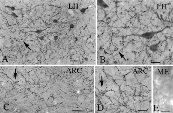
A new orexigenic peptide called hypocretin (orexin) has recently been described in neurons of the lateral hypothalamus and perifornical area. The medial and lateral hypothalamus have been loosely called satiety and feeding centers of the brain, respectively. Approximately one-third of all medial and lateral hypothalamic neurons tested, but not hippocampal neurons, show a striking nanomolar sensitivity to hypocretin. As studied with calcium digital imaging with fura-2, hypocretin raises cytoplasmic calcium via a mechanism based on G-protein enhancement of calcium influx through plasma membrane channels. The peptide has a potent effect at both presynaptic and postsynaptic receptors. Most synaptic activity in hypothalamic circuits is attributable to axonal release of GABA or glutamate. With whole-cell patch-clamp recording, we show that hypocretin, acting directly at axon terminals, can increase the release of each of these amino acid transmitters. Two hypocretin peptides, hypocretin-1 and hypocretin-2, are coded by a single gene; neurons that respond to one peptide also respond to the other. In addition to its effect on feeding, we find that this peptide also regulates the synaptic activity of physiologically identified neuroendocrine neurons studied in hypothalamic slices containing the arcuate nucleus, suggesting a second function of hypocretin in hormone regulation. The widespread distribution of hypocretin axons, coupled with the strong response to the peptide at both presynaptic and postsynaptic sites, suggests that the peptide probably modulates a variety of hypothalamic regulatory systems and could regulate the axonal input to these regions presynaptically.


Figures

Fig. 1.
Fig. 1.
Both excitatory and inhibitory neurons express hypocretin receptors. To determine the transmitter identity of neurons that respond to hypocretin, different transmitter antagonists were used, and responses to hypocretin were studied with whole-cell recording in voltage-clamped cells held at −60 mV. A, In the presence of glutamate receptor antagonists AP-5 (100 μm) and CNQX (10 μm), only sIPSPs were found, and these were completely blocked with the GABAAantagonist bicuculline (20 μm) (data not shown). When hypocretin (1 μm) was bath-applied, the frequency of sIPSCs increased dramatically, and after peptide washout, the frequency of sIPSCs decreased to its normal baseline. B, In the presence of the GABAA antagonist bicuculline (20 μm), sEPSCs were found, and these were completely blocked by AP-5 (100 μm) and CNQX (10 μm) (data not shown). Hypocretin (1 μm) increased the frequency of sEPSCs, and these returned to normal after peptide washout. Both GABA and glutamate evoked inward currents (downward deflection) attributable to the composition of the pipette solution. C,D, Mean increase in sEPSCs and sIPSCs evoked by hypocretin and the return toward control levels after peptide washout.
Fig. 2.
Fig. 2.
Hypocretin immunoreactivity. A, Immunocytochemical staining revealed high densities of axons in both the lateral hypothalamus (A, B,LH) and arcuate nucleus (C,D, ARC) of the hypothalamus. Around immunoreactive cells in the LH, a high density of immunoreactive axons was found. E, Immunoreactive axons were not found in the external zone of the median eminence (ME), in which neurosecretory axons release pituitary tropins that are carried to the anterior pituitary by the portal blood system. Scale bars:A, 25 μm; B, 12 μm; C, 25 μm; D, E, 12 μm.
Fig. 3.
Fig. 3.
Hypothalamic slice electrophysiology.A, Experimental paradigm for recording hypothalamic slices that included the arcuate nucleus (ARC).B, In the presence of AP-5 (100 μm) and CNQX (10 μm), IPSPs were evoked by orthodromic electrical stimulation (asterisk), as shown in A. The IPSP was blocked in the presence of bicuculline (20 μm), indicating that it was attributable to GABA release. Hypocretin (HCRT) applied by microdrop (30 μm) caused a substantial increase in the amplitude of the IPSP. This recovered to baseline levels after washout of the peptide. Each trace is the mean of eight sweeps. C, After testing hypocretin in B, the axon of the same cell was antidromically stimulated from the median eminence. That median eminence stimulation evoked an action potential even in the presence of control application of Co2+ (1 mm) indicated that the action potential response was not attributable to recurrent collaterals releasing transmitters on the recorded cell. A fixed latency of 3–5 mS between stimulus artifact and antidromic spike was routinely found. These data indicate that hypocretin enhances the action of transmitters released onto identified neuroendocrine neurons.
Fig. 4.
Fig. 4.
Calcium responses to hypocretin. A, Based on fura-2 calcium recordings, neurons from the lateral hypothalamus (LH), the site of the hypocretin immunoreactive cell bodies, showed strong responses to hypocretin (HCRT) (1 μm) and clear recovery after two bath applications of the peptide. Horizontal lines above Ca2+ trace indicate time of drug application. Except for Figure 3C, all other experiments used hypocretin-2. B, Medial hypothalamus cells also showed strong Ca2+ elevations in response to hypocretin but no response to the C-terminal 1–17 (1–17) peptide of preprohypocretin (1 μm). C, Hypocretin-1 (HCRT: 1), using the 33 amino acid structure with the double disulfide bonds found for orexin A, and hypocretin-2 (HCRT: 2) were compared. Each evoked a Ca2+ rise in this typical medial hypothalamic neuron. D, Hypothalamic neuron that showed an increasing peak response to increasing concentrations of the peptide, with no response to 1 nm hypocretin, a response to 10 nm hypocretin, and a bigger response to 100 nmhypocretin.
Fig. 5.
Fig. 5.
Mechanisms of hypocretin elevation of calcium.A, To demonstrate that hypocretin can evoke Ca2+ elevations directly at the cell body rather than by altering synaptic interactions, hypocretin (1 μm) was added in the presence of tetrodotoxin (TTX) (1 μm), used to block action potential-mediated release of transmitters. B, Depleting intracellular stores of Ca2+ with thapsigargin (2 μm) pretreatment did not affect the action of hypocretin in elevating Ca2+ in the presence of 1 μm TTX.C, Eliminating extracellular Ca2+ and adding the Ca2+ chelator EGTA (1 mm) completely blocked the Ca2+ rise evoked by 1 μm hypocretin (HCRT). When bath Ca2+ levels were returned to normal, the action of hypocretin returned. D, Cd2+ (100 μm) blocked the Ca2+ rise, indicating an extracellular origin of Ca2+. E, Pretreatment of hypothalamic neurons with bisindolylmaleide (1 μm for 12 hr), a PKC inhibitor, completely blocked the actions of hypocretin (1 μm). Glutamate (GLU) (10 μm), applied as a control, evoked a large Ca2+ rise. F, Most cortical cells showed no response to hypocretin, as shown by the typical cell in f-1. A small percent of cortical cells did show a small Ca2+ rise in response to hypocretin, as shown in f-2. A control application of glutamate (GLU) (10 μm) evoked a Ca2+ rise.
Fig. 6.
Fig. 6.
Presynaptic mechanism for hypocretin to enhance GABA or glutamate secretion. In the presence of TTX (1 μm), mPSCs were studied with whole-cell voltage-clamp recording. A, In the presence of the glutamate receptor antagonists AP-5 (100 μm) and CNQX (10 μm), hypocretin (1 μm) increased the frequency of mIPSCs. After washout of the peptide, the frequency of mIPSCs returned to baseline levels. B, An example of hypocretin (1 μm) increasing the frequency of mEPSCs in the presence of bicuculline (20 μm). C,D, Mean increase in mEPSCs and mIPSCs and the return toward baseline levels after peptide washout. E, Cumulative probability for mEPSC amplitude in the presence of hypocretin (1 μm) or in its absence (control). Superimposition of the two lines indicates that although hypocretin increased the frequency of mEPSCs, it did not change the amplitude, suggesting that the peptide acted at a presynaptic site to enhance transmitter release.

References

    1. Belousov AB, van den Pol AN. Local synaptic release of glutamate from neurons in the rat hypothalamic arcuate nucleus. J Physiol (Lond) 1997;499:747–761. - PMC - PubMed
    1. Bleakman D, Harrison NL, Colmers WF, Miller RJ. Investigations into neuropeptide Y-mediated presynaptic inhibition in cultured hippocampal neurones of the rat. Br J Pharmacol. 1992;107:334–340. - PMC - PubMed
    1. Chen G, van den Pol AN. NPY Y1- and Y2-like receptors coexist in pre- and postsynaptic sites: inhibition of GABA release in isolated self-innervating SCN neurons. J Neurosci. 1996;16:7711–7724. - PMC - PubMed
    1. Chen G, van den Pol AN. Presynaptic GABA-B autoreceptor modulation of P/Q type calcium channels and GABA release in rat suprachiasmatic nucleus neurons. J Neurosci. 1998;18:1913–1922. - PMC - PubMed
    1. Christian EP, Dudek F. Characteristics of local excitatory circuits studied with glutamate microapplication in the CA3 area of rat hippocampal slices. J Neurophysiol. 1988;59:90–109. - PubMed

Publication types

MeSH terms