. 1998 Sep 21;188(6):1029-37.
doi: 10.1084/jem.188.6.1029.

Urokinase receptor (CD87) regulates leukocyte recruitment via beta 2 integrins in vivo

Affiliations

Urokinase receptor (CD87) regulates leukocyte recruitment via beta 2 integrins in vivo

A E May et al. J Exp Med. .

Abstract

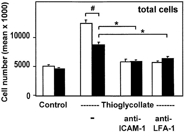
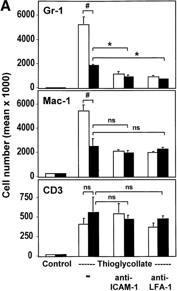
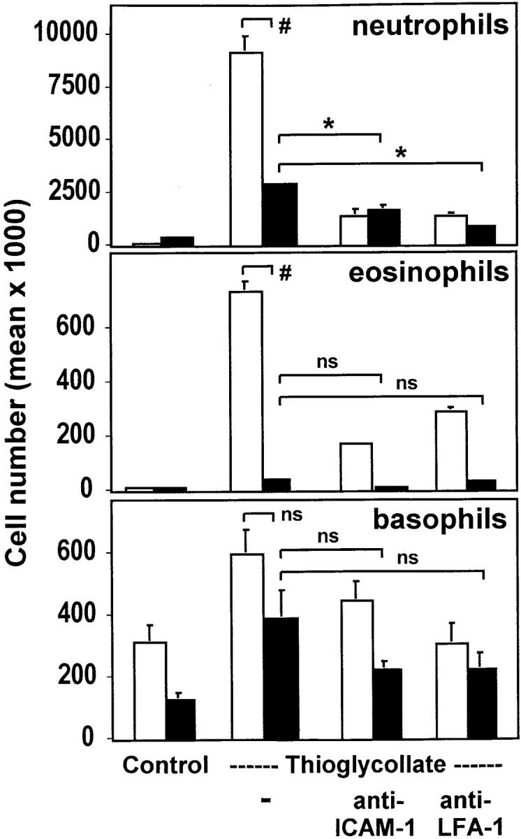
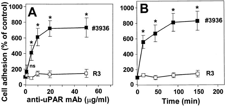
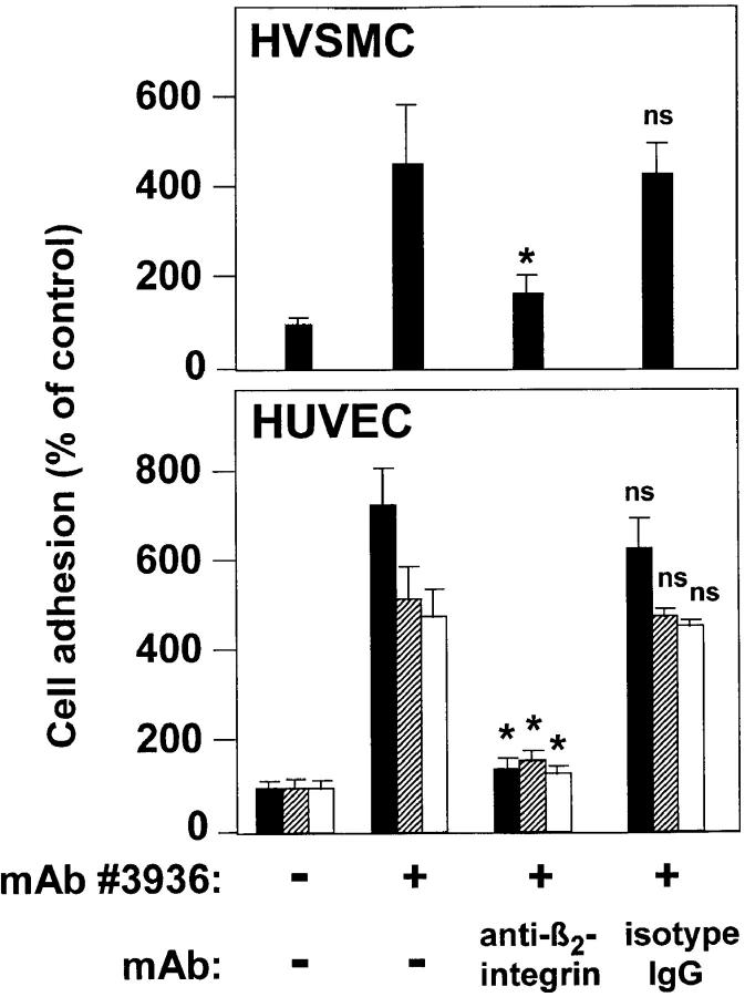
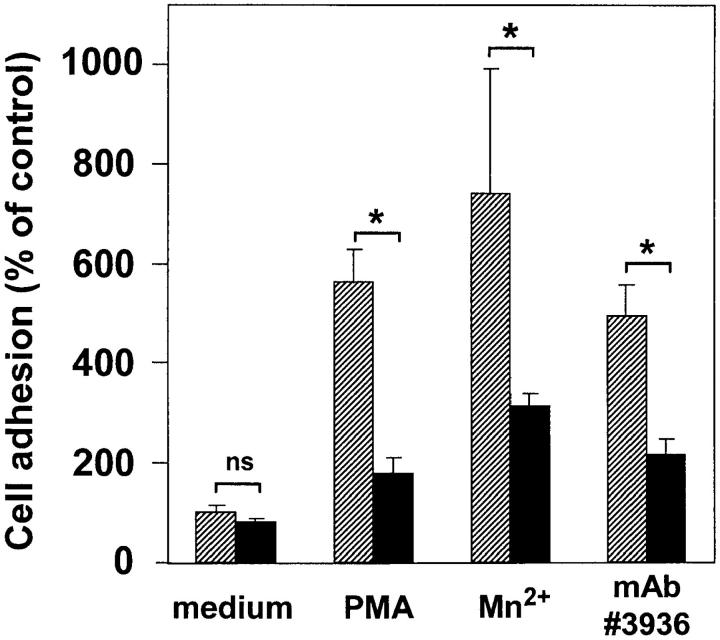
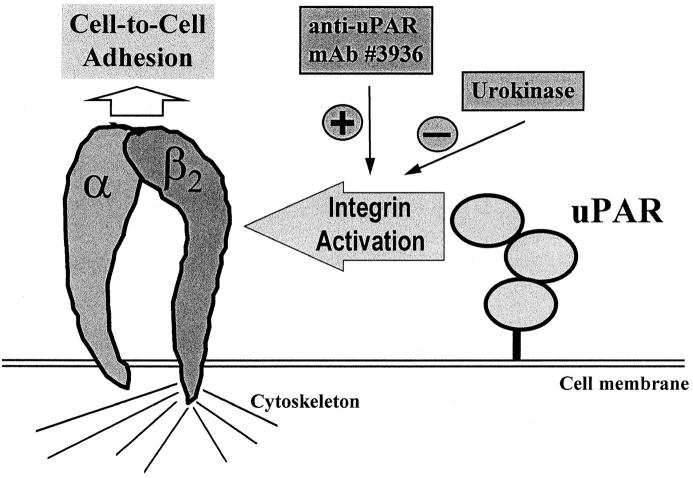
The urokinase receptor (CD87; uPAR) is found in close association with beta 2 integrins on leukocytes. We studied the functional consequence of this association for leukocyte adhesion and migration. In vivo, the beta 2 integrin-dependent recruitment of leukocytes to the inflamed peritoneum of uPAR-deficient mice was significantly reduced as compared with wild-type animals. In vitro, beta 2 integrin-mediated adhesion of leukocytes to endothelium was lost upon removal of uPAR from the leukocyte surface by phosphatidyl-inositol-specific phospholipase C. Leukocyte adhesion was reconstituted when soluble intact uPAR, but not a truncated form lacking the uPA-binding domain, was allowed to reassociate with the cell surface. uPAR ligation with a monoclonal antibody induced adhesion of monocytic cells and neutrophils to vascular endothelium by six- to eightfold, whereas ligation with inactivated uPA significantly reduced cell-to-cell adhesion irrespective of the beta 2 integrin-stimulating pathway. These data indicate that beta 2 integrin-mediated leukocyte-endothelial cell interactions and recruitment to inflamed areas require the presence of uPAR and define a new phenotype for uPAR-deficient mice. Moreover, uPAR ligation differentially modulates leukocyte adhesion to endothelium and provides novel targets for therapeutic strategies in inflammation-related vascular pathologies.


Figures

Figure 1
Figure 1
Leukocyte emigration in thioglycollate-induced peritonitis. Wild-type mice (white bars) and uPAR-deficient mice (black bars) were injected intraperitoneally with buffer alone (Control) or with thioglycollate solution in the absence or presence of the indicated antibodies. After leukocyte emigration for 4 h, mice were killed and the leukocytes were counted in lavages from the peritoneum. SE was calculated from four animals for each condition and the experiment was repeated twice. # P < 0.02; *P < 0.01.
Figure 2
Figure 2
Analysis of subpopulations of emigrated leukocytes in the peritoneal lavage. Leukocytes obtained in peritoneal lavages after induction of peritonitis for 4 (A) or 24 h (B) from wild-type mice (white bars) and uPAR-deficient mice (black bars) were analyzed by flow cytometry for the expression of Gr-1 (anti-granulocytes), Mac-1 (anti-myeloid cells), or CD3 (anti-CD3 TCR complex). Absolute numbers of cells were calculated from the percentage of stained cells and the number of total emigrated cells shown in Fig. 1. # P < 0.01; *P < 0.002; § P < 0.005.
Figure 2
Figure 2
Analysis of subpopulations of emigrated leukocytes in the peritoneal lavage. Leukocytes obtained in peritoneal lavages after induction of peritonitis for 4 (A) or 24 h (B) from wild-type mice (white bars) and uPAR-deficient mice (black bars) were analyzed by flow cytometry for the expression of Gr-1 (anti-granulocytes), Mac-1 (anti-myeloid cells), or CD3 (anti-CD3 TCR complex). Absolute numbers of cells were calculated from the percentage of stained cells and the number of total emigrated cells shown in Fig. 1. # P < 0.01; *P < 0.002; § P < 0.005.
Figure 3
Figure 3
Quantitation of granulocyte subpopulations in the peritoneal lavage. The migrated leukocytes from wild-type mice (white bars) and uPAR-deficient mice (black bars) were cytocentrifuged and stained with May-Grünwald-Giemsa solution. The quantitation of cell numbers of leukocyte subpopulations was performed by light microscopy. SE was calculated from four animals for each condition and the experiment was repeated twice. # P < 0.02; *P < 0.005.
Figure 4
Figure 4
Requirement of uPAR for leukocyte adhesion to endothelial cells. Myelo-monocytic HL60 cells were either not treated (hatched bars) or pretreated with piPLC (0.5 U/ml, 90 min, 37°C; black bars), washed, and incubated for 10 min without or with soluble intact uPAR (0.8 μg/ml), followed by stimulation for 20 min with (A) PMA (10 ng/ml) or (B) Mn2+ (1 mM). After another washing step, the adhesion assay was performed in the absence or presence of anti–β2 integrin mAb 60.3 (10 μg/ml). Values are displayed as percent of PMA- or Mn2+-inducible adhesion and represent the mean ± SEM of three independent experiments. *P < 0.001 as compared with control.
Figure 5
Figure 5
Reconstitution of leukocyte adhesion to endothelium by soluble uPAR. Adhesion of monocytic cells to HUVECs was performed as described in the legend to Fig. 4. piPLC-pretreated myelo-monocytic HL60 cells were incubated with increasing concentrations of soluble intact (D1/D2/D3) uPAR or with its truncated form (D2/D3) lacking domain 1, respectively, and were subsequently stimulated by PMA. Values are displayed as percentage of PMA-inducible adhesion in non–piPLC-pretreated cells and one representative experiment out of three is shown. # P < 0.01 and *P < 0.001 as compared with piPLC-pretreated cells without soluble uPAR.
Figure 6
Figure 6
Effect of different anti-uPAR mAbs on leukocytic cell adhesion to endothelium. Myelo-monocytic HL60 cells were incubated for 30 min with different anti-uPAR mAbs (20 μg/ml each) representing uPA-blocking (no. 3936, R3, R9) or -nonblocking (R2, R4, R8) activity, with (Fab′)2 of mAb no. 3936 (12 μg/ml), boiled (5 min) mAb no. 3936, or control IgG2a as indicated. After washing, the adhesion assay was performed. Values are displayed as percentage of control (no antibody added) and represent the mean (± SEM) of at least three independent experiments. *P < 0.001 as compared with control.
Figure 7
Figure 7
Induction of leukocyte adhesion to endothelium by anti-uPAR mAb no. 3936. Anti-uPAR mAb no. 3936 (▪) or R3 (□) were added to myelo-monocytic HL60 cells for 1 h at different concentrations (A), or at a concentration of 10 μg/ml for different time intervals (B). After washing, the adhesion assay was performed as described. Values (mean ± SEM, n = 3) are displayed as percentage of control (no antibody added). One representative experiment out of three is shown. *P < 0.001 as compared with control (no antibody added or time 0).
Figure 8
Figure 8
Induction of leukocyte adhesion to endothelial and smooth muscle cells by anti-uPAR mAb no. 3936. Myelo-monocytic HL60 cells (black bars), monocytic U937 cells (hatched bars) and freshly isolated neutrophils (white bars) were pretreated with anti-uPAR mAb no. 3936 (20 μg/ml) for 30 min. After washing, the adhesion assay was performed in the absence or presence of anti–β2 integrin mAb 60.3 or isotype control mAb (10 μg/ml) as indicated. Values (mean ± SEM, n = 3) are displayed as percentage of control (no antibody added), and one representative experiment out of three is shown. *P < 0.001 as compared with anti-uPAR-induced adhesion.
Figure 9
Figure 9
Interference of uPA with leukocyte adhesion to endothelium. Myelo-monocytic HL60 cells were incubated in the absence (hatched bars) or the presence (black bars) of inactivated uPA (50 nM) for 30 min (37°C, 5% CO2) before addition of medium, PMA (10 ng/ml), Mn2+ (1.0 mM), or anti-uPAR mAb no. 3936 (10 μg/ml) for another 30 min, as indicated. After washing, the adhesion assay was performed. Values are displayed as percent of untreated control (medium) and represent the mean ± SEM of three independent experiments. *P < 0.01.
Figure 10
Figure 10
Hypothetical model for the uPAR–β2 integrin cross-talk. The presence of uPAR on leukocyte subpopulations is required for their adhesion to endothelium and their subsequent recruitment into the inflamed tissue as documented here for a peritonitis model. In vitro, ligation of uPAR by a specific mAb no. 3936 induces leukocyte adhesion to endothelial cells, whereas uPA greatly inhibits cell-to-cell interactions independent of the stimulatory pathway.

Similar articles

Cited by

References

    1. Springer TA. Traffic signals for lymphocyte recirculation and leukocyte emigration: the multistep paradigm. Cell. 1994;76:301–314. - PubMed
    1. Carlos TM, Harlan JM. Leukocyte-endothelial adhesion molecules. Blood. 1994;84:2068–2101. - PubMed
    1. Larson RS, Springer TA. Structure and function of leukocyte integrins. Immunol Rev. 1990;114:181–217. - PubMed
    1. Van der Vieren M, Le Trong H, Wood CL, Moore PF, St. John T, Staunton DE, Gallatin WM. A novel leukointegrin, αdβ2, binds preferentially to ICAM-3. Immunity. 1995;3:683–690. - PubMed
    1. Smith CW, Marlin SD, Rothlein R, Toman C, Anderson DC. Cooperative interactions of LFA-1 and Mac-1 with intercellular adhesion molecule-1 in facilitating adherence and transendothelial migration of human neutrophils in vitro. J Clin Invest. 1989;83:2008–2017. - PMC - PubMed

Publication types

MeSH terms