. 1998 Sep 21;188(6):1039-46.
doi: 10.1084/jem.188.6.1039.

Inhibition of angiogenesis by interleukin 4

Affiliations

Inhibition of angiogenesis by interleukin 4

O V Volpert et al. J Exp Med. .

Abstract

Interleukin (IL)-4, a crucial modulator of the immune system and an active antitumor agent, is also a potent inhibitor of angiogenesis. When incorporated at concentrations of 10 ng/ml or more into pellets implanted into the rat cornea or when delivered systemically to the mouse by intraperitoneal injection, IL-4 blocked the induction of corneal neovascularization by basic fibroblast growth factor. IL-4 as well as IL-13 inhibited the migration of cultured bovine or human microvascular cells, showing unusual dose-response curves that were sharply stimulatory at a concentration of 0.01 ng/ml but inhibitory over a wide range of higher concentrations. Recombinant cytokine from mouse and from human worked equally well in vitro on bovine and human endothelial cells and in vivo in the rat, showing no species specificity. IL-4 was secreted at inhibitory levels by activated murine T helper (TH0) cells and by a line of carcinoma cells whose tumorigenicity is known to be inhibited by IL-4. Its ability to cause media conditioned by these cells to be antiangiogenic suggested that the antiangiogenic activity of IL-4 may play a role in normal physiology and contribute significantly to its demonstrated antitumor activity.


Figures

Figure 1
Figure 1
Local IL-4 blocks neovascularization in vivo in the rat cornea. Hydron pellets containing bFGF at 100 ng/ml, recombinant murine IL-4 at 100 ng/ml or the combination with or without anti–IL-4 antibody at 40 μg/ml were implanted into the avascular cornea of the rat. 7 d later vessels were filled with colloidal carbon and corneas photographed. Note the vigorous angiogenic response to bFGF and its inhibition by IL-4.
Figure 2
Figure 2
The biphasic, serum-dependent effect of recombinant IL-4 on the migration of capillary endothelial cells. (A) Serum-starved bovine adrenal capillary endothelial cells were allowed to migrate towards murine IL-4 in the absence (circles) or in the presence (triangles) of an inducing concentration of bFGF. Identical results were obtained using recombinant human IL-4. (B) IL-13 tested as was IL-4 in A. (C) Serum-starved human dermal capillary endothelial cells were allowed to migrate towards murine IL-4 (open symbols) or towards human IL-4 (filled symbols) in the absence (circles) or presence (triangles) of bFGF. Note: In all graphs, dotted lines indicate migration seen towards vehicle (0.1% bovine serum albumin, BSA) or towards 10 ng/ml bFGF (bFGF); bars indicate SE.
Figure 2
Figure 2
The biphasic, serum-dependent effect of recombinant IL-4 on the migration of capillary endothelial cells. (A) Serum-starved bovine adrenal capillary endothelial cells were allowed to migrate towards murine IL-4 in the absence (circles) or in the presence (triangles) of an inducing concentration of bFGF. Identical results were obtained using recombinant human IL-4. (B) IL-13 tested as was IL-4 in A. (C) Serum-starved human dermal capillary endothelial cells were allowed to migrate towards murine IL-4 (open symbols) or towards human IL-4 (filled symbols) in the absence (circles) or presence (triangles) of bFGF. Note: In all graphs, dotted lines indicate migration seen towards vehicle (0.1% bovine serum albumin, BSA) or towards 10 ng/ml bFGF (bFGF); bars indicate SE.
Figure 3
Figure 3
Effect of IL-4 on the proliferation of human capillary endothelial cells. Human dermal microvascular endothelial cells were allowed to grow over 72 h in the presence of increasing concentrations of IL-4 (open circles) or the combination of IL-4 and 100 ng/ml bFGF (filled circles) and proliferation quantitated. Note: Dotted lines indicate growth in the absence of any cytokine additions (no proliferation) and growth in the presence of 100 ng/ml bFGF (bFGF). Similar results were obtained if serum was used instead of bFGF or HUVEC cells instead of HMVECs. Bars indicate SE.
Figure 4
Figure 4
IL-4 is responsible for the lack of in vitro angiogenic activity in revertant K485 cells. (A) Media conditioned by K485 carcinoma cells transfected with vector (F1-1) or transfected with murine IL-4 and expressing the cytokine at low levels (E2A5, E2A6) and at high levels (D2B1) were tested for ability to induce the migration of bovine capillary endothelial cells alone, when mixed with bFGF (10 ng/ml) and/or with neutralizing anti–muIL-4 antibodies (32 μl/ml). Controls labeled DME include tests of antibodies alone (AB) and of murine IL-4 with other components as indicated. Note: dotted lines as in legend to Fig. 1. (B) Supernatants of vector-transfected line F1-1 and IL-4– transfected revertant D2B1 were mixed 1:1 and tested for the ability to induce the migration of bovine capillary endothelial cells. (C) Media conditioned by parental F1-1 and revertant D2B1 cells was incorporated into pellets with the indicated additions and tested in the rat cornea for ability to induce neovascularization. An asterisk indicates significant difference from parental F1-1 line by unpaired Student's t test, P < 0.002.
Figure 4
Figure 4
IL-4 is responsible for the lack of in vitro angiogenic activity in revertant K485 cells. (A) Media conditioned by K485 carcinoma cells transfected with vector (F1-1) or transfected with murine IL-4 and expressing the cytokine at low levels (E2A5, E2A6) and at high levels (D2B1) were tested for ability to induce the migration of bovine capillary endothelial cells alone, when mixed with bFGF (10 ng/ml) and/or with neutralizing anti–muIL-4 antibodies (32 μl/ml). Controls labeled DME include tests of antibodies alone (AB) and of murine IL-4 with other components as indicated. Note: dotted lines as in legend to Fig. 1. (B) Supernatants of vector-transfected line F1-1 and IL-4– transfected revertant D2B1 were mixed 1:1 and tested for the ability to induce the migration of bovine capillary endothelial cells. (C) Media conditioned by parental F1-1 and revertant D2B1 cells was incorporated into pellets with the indicated additions and tested in the rat cornea for ability to induce neovascularization. An asterisk indicates significant difference from parental F1-1 line by unpaired Student's t test, P < 0.002.
Figure 5
Figure 5
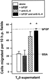
Supernatants of T0 cells are antiangiogenic due to IL-4. Supernatants obtained from splenic cells freshly derived from D011.10 T cell receptor transgenic mice and incubated with ovalbumin for 72 h were tested for the ability to induce the migration of bovine capillary endothelial cells alone and with either bFGF, anti–IL-4 or both. Dotted lines indicate migration of cells towards bFGF alone or towards media alone (BSA). An asterisk indicates significant difference from antibody-free control, P < 0.005.

Similar articles

Cited by

References

    1. Paul WE. Interleukin-4: a prototypic immunoregulatory lymphokine. Blood. 1991;77:1859–1870. - PubMed
    1. Kopf M, Le Gros G, Bachmann M, Lamers KC, Bluethmann H, Kohler G. Disruption of the murine IL-4 gene blocks Th2 cytokine responses. Nature. 1993;362:245–248. - PubMed
    1. Kuhn R, Rajewsky K, Muller W. Generation and analysis of interleukin-4 deficient mice. Science. 1991;254:707–710. - PubMed
    1. Ohara J, Paul WE. Receptors for B-cell stimulatory factor-1 expressed on cells of haematopoietic lineage. Nature. 1987;325:537–540. - PubMed
    1. Mosmann TR, Yokota T, Kastelein R, Zurawski SM, Arai N, Takebe Y. Species-specificity of T cell stimulating activities of IL 2 and BSF-1 (IL-4): comparison of normal and recombinant, mouse and human IL-2 and BSF-1 (IL-4) J Immunology. 1987;138:1813–1816. - PubMed

Publication types

MeSH terms