. 1998 Sep 21;188(6):1105-16.
doi: 10.1084/jem.188.6.1105.

Major histocompatibility complex class I viral antigen processing in the secretory pathway defined by the trans-Golgi network protease furin

Affiliations

Major histocompatibility complex class I viral antigen processing in the secretory pathway defined by the trans-Golgi network protease furin

B C Gil-Torregrosa et al. J Exp Med. .

Abstract

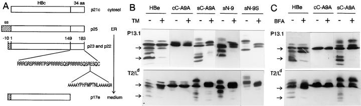
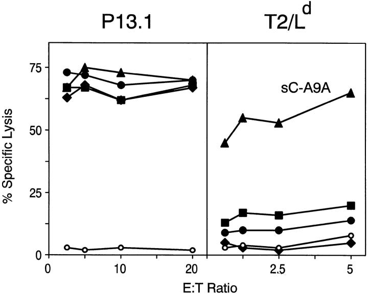
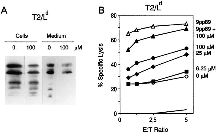
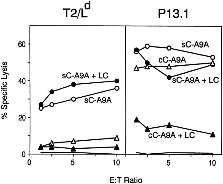
Classical antigen presentation by major histocompatibility complex class I molecules involves cytosolic processing of endogenously synthesized antigens by proteasomes and translocation of processed peptides into the endoplasmic reticulum (ER) by transporters associated with antigen presentation (TAP). Alternative pathways for processing of endogenous antigens, generally involving the ER, have been suggested but not fully proved. We analyzed the potential for class I presentation of proteolytic maturation of secretory antigens in the exocytic pathway. We found that hepatitis B (HB) virus secretory core protein HBe can efficiently deliver COOH-terminally located antigenic peptides for endogenous class I loading in the absence of TAP. Antigen presentation to specific cytotoxic T lymphocytes correlates with protein maturation at the COOH terminus, since modification of maturation and transport of HBe through the secretory pathway alters antigen presentation. Both maturation and a necessary processing step occur in the Golgi or post-Golgi compartment. Antigen presentation is independent of proteasome activity, but inhibitors of the trans-Golgi network resident protease furin inhibit both HBe maturation and antigen presentation. These results define a new antigen processing pathway located in the secretory route, with a central role for proteolytic maturation mediated by the subtilisin protease family member furin as an efficient source for antigen presentation.


Figures

Figure 3
Figure 3
Maturation of HBe and derived chimeric proteins. (A) Schematic representation of main HBe and HBc forms during the maturation process. Localization of 9pp89 epitope (YPHFMPTNL) with flanking residues in sC-A9A construct is shown. aa, Amino acids. ss, Signal sequence. (B) Western blot analysis of HBe-derived chimeras in rVV-infected cells. Complete cellular extracts of rVV-infected P13 cells and rVV-infected T2/Ld cells in the presence or absence of tunicamycin (TM) or BFA (C) were separated by SDS-PAGE, transferred to a membrane, and detected with an anti-HBe antiserum. Arrows, Positions of p22 and p17 HBe forms.
Figure 1
Figure 1
TAP-independent presentation to CTLs of secreted chimeric HBe proteins. P13 TAP+ or T2/Ld TAP cells infected with rVV encoding sC-A9A (filled triangles), cC-A9A (filled circles), sN-9 (filled diamonds), eN-A9A (filled squares), or HBe (open circles) were confronted in a 51Cr- release assay with 9pp89-specific CTLs restricted by H2-Ld. See Materials and Methods for rVV nomenclature.
Figure 2
Figure 2
TAP-independent recognition of 9pp89 by CTLs requires endogenous processing of sC-A9A chimeric protein. (A) 51Cr-labeled T2/Ld target cells infected with either rVV–sC-A9A or rVV-HBe or mock-infected were incubated with 40 nM 9pp89 peptide, supernatant from rVV–sC-A9A– infected T2/Ld cells, or cocultured with rVV-infected cold cells as indicated, and tested in a CTL assay. Recognition of rVV-HBe– and rVV–sC-A9A–infected controls without further treatment and of rVV–sC-A9A–infected targets immediately after infection (0 h) are shown. Specific lysis at an E/T ratio of 5:1 is shown. (B) Effect of BFA (filled symbols) on recognition of 9pp89 peptide– loaded (open triangles) and rVV–sC-A9A– infected (open circles) T2/Ld cells by specific CTLs. The negative control (solid line) was provided by rVV-HBe–infected T2/Ld cells.
Figure 4
Figure 4
Effect on maturation and antigen presentation of alteration of the secretory pathway by pepstatin. (A) Cell extracts and supernatant culture medium of T2/Ld cells infected with rVV–sC-A9A and treated with the indicated amounts of the aspartic protease inhibitor pepstatin were analyzed by Western blot with HBe-specific antiserum. (B) CTL recognition of T2/Ld cells infected with rVV–sC-A9A (open circles) and treated with 100 μM (filled circles), 25 μM (filled diamonds), and 6.25 μM (filled squares) pepstatin. A control of 9pp89 peptide–loaded target cells (filled triangles) treated or untreated (open triangles) with pepstatin is included. The negative control (solid line) was provided by rVV-HBe–infected T2/Ld cells.
Figure 5
Figure 5
Effect of the proteasome inhibitor lactacystin (LC) on antigen presentation of HBe-derived chimeric proteins. Recognition by 9pp89-specific CTLs of cell lines T2/Ld and P13.1 infected with rVV– sC-A9A (open circles) or rVV–cC-A9A (open triangles) treated with lactacystin (filled symbols) 10 μM in the case of T2/Ld cells, 30 μM for rVV–cC-A9A–infected P13.1, and 100 μM for rVV–sC-A9A–infected P13.1 cells is shown. The negative control (solid line) was provided by rVV-HBe– infected cells.
Figure 6
Figure 6
Furin inhibitor decRVKR-CMK inhibits maturation and antigen presentation of sC-A9A protein. (A) CTL recognition of rVV– sC-A9A–infected T2/Ld cells untreated (open circles) or treated with furin inhibitor (filled symbols) at the indicated concentrations. 8 μM* (filled circles) signifies that a second addition of 8 μM decRVKR-CMK was performed before CTL assay. The negative control (solid line) was provided by rVV-HBe–infected T2/Ld cells. (B) Cell extracts from rVV–sC-A9A– infected T2/Ld and P13.1 cells treated with the indicated amounts of decRVKR-CMK during infection were analyzed by Western blot with an anti-HBe antiserum. In the 8* lane, a second addition of 8 μM decRVKR-CMK was performed 3 h before cell harvesting.
Figure 7
Figure 7
Characterization and quantitation of naturally processed antigenic peptides in cell extracts. Acid-soluble extracts prepared from T2/Ld (filled circles), T2 (filled diamonds), L/Ld (filled squares), or Ltk (filled triangles) cells infected with rVV–sC-A9A (filled symbols) or rVV–cC-A9A (open symbols) and fractionated by reverse-phase HPLC were tested for their ability to sensitize P13 cells for lysis by 9pp89-specific CTLs. (A) Detection of peptides by 9pp89-specific CTLs after HPLC fractionation of whole cell extracts. Each test well contained 9 × 10⁶ cell equivalents. (B) Peptide quantitation by CTL recognition of serial dilutions of antigenic fractions. The average of specific lysis of fractions 62 and 69, the peaks of the two CTL-reactive peptide forms, is shown. 10% average specific lysis was chosen as the minimum level for considering positive T cell recognition and comparison among different rVV and lines.

Similar articles

Cited by

References

    1. Matsumura M, Fremont DH, Peterson PA, Wilson IA. Emerging principles for the recognition of peptide antigens by MHC class I molecules. Science. 1992;257:927–934. - PubMed
    1. Bouvier M, Wiley DC. Importance of peptide amino and carboxyl termini to the stability of MHC class I molecules. Science. 1994;265:398–402. - PubMed
    1. Del Val M, Hengel H, Hacker H, Hatlaub U, Ruppert T, Lucin P, Koszinowski UH. Cytomegalovirus prevents antigen presentation by blocking the transport of peptide-loaded major histocompatibility complex class I molecules into the medial-Golgi compartment. J Exp Med. 1992;176:729–738. - PMC - PubMed
    1. York IA, Rock KL. Antigen processing and presentation by the class I major histocompatibility complex. Annu Rev Immunol. 1996;14:369–396. - PubMed
    1. Coux O, Tanaka K, Goldberg AL. Structure and functions of the 20S and 26S proteasome. Annu Rev Biochem. 1996;65:801–847. - PubMed

Publication types

MeSH terms