. 1998 Sep 21;188(6):1117-24.
doi: 10.1084/jem.188.6.1117.

Asymmetric cell divisions sustain long-term hematopoiesis from single-sorted human fetal liver cells

Affiliations

Asymmetric cell divisions sustain long-term hematopoiesis from single-sorted human fetal liver cells

T H Brummendorf et al. J Exp Med. .

Abstract

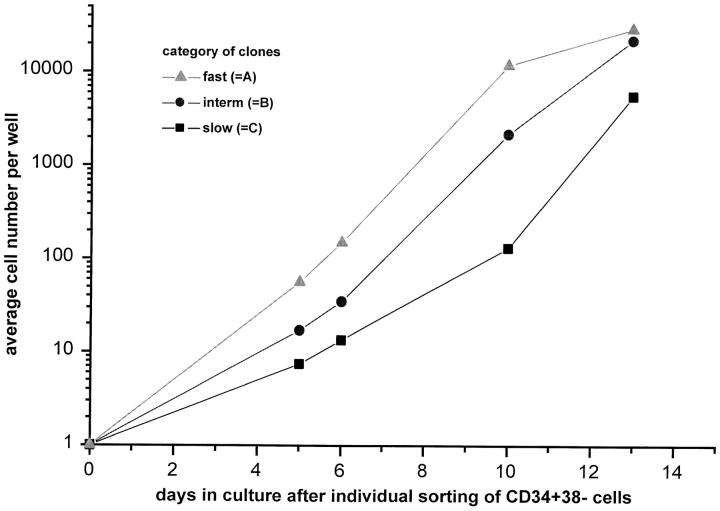
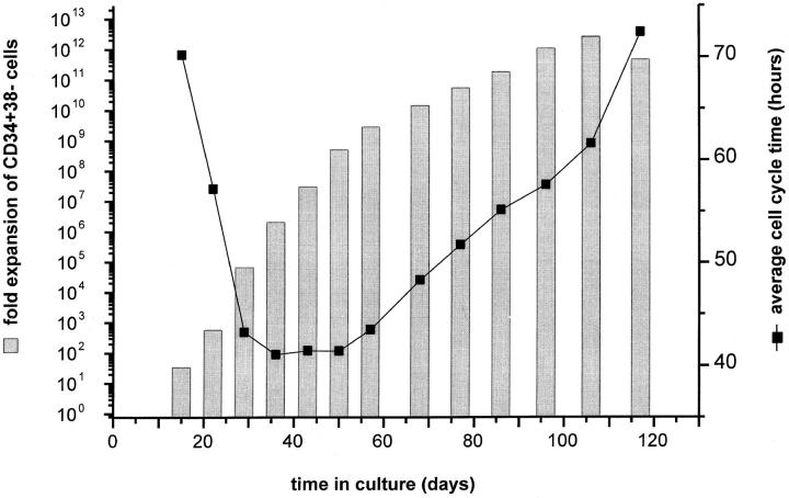
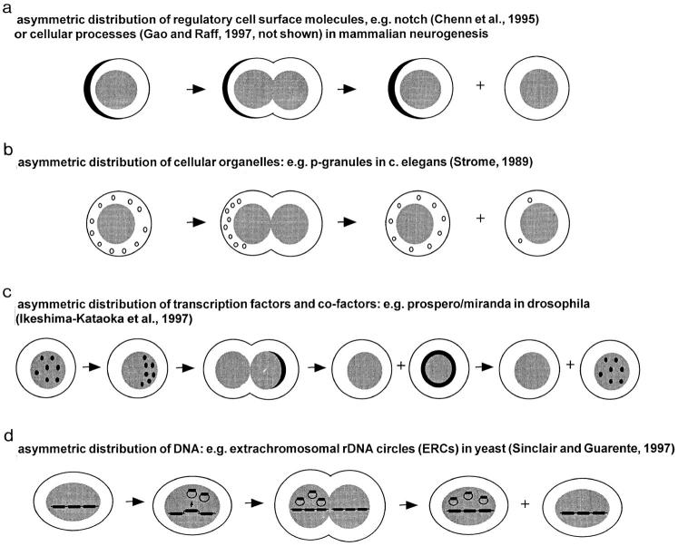
Hematopoietic stem cells (HSCs) in adult marrow are believed to be derived from fetal liver precursors. To study cell kinetics involved in long-term hematopoiesis, we studied single-sorted candidate HSCs from fetal liver that were cultured in the presence of a mixture of stimulatory cytokines. After 8-10 d, the number of cells in primary cultures varied from <100 to >10,000 cells. Single cells in slow growing colonies were recloned upon reaching a 100-200 cell stage. Strikingly, the number of cells in subclones varied widely again. These results are indicative of asymmetric divisions in primitive hematopoietic cells in which proliferative potential and cell cycle properties are unevenly distributed among daughter cells. The continuous generation of functional heterogeneity among the clonal progeny of HSCs is in support of intrinsic control of stem cell fate and provides a model for the long-term maintenance of hematopoiesis in vitro and in vivo.


Figures

Figure 1
Figure 1
FACS® profile of the CD34+CD38 candidate HSCs sorted from fetal liver to initiate single cell cultures in 96-well plates. Viable, propidium iodide negative (PI) CD34+CD38 cells with a low side scatter (SSC) and low levels of CD45RA and CD71 expression were recovered from the indicated sort windows.
Figure 2
Figure 2
Experimental design of study.
Figure 3
Figure 3
Clonal heterogeneity in early proliferative response of single-sorted CD34+CD38 fetal liver cells cultured in growth factor–supplemented SFM. Single CD34+CD38 fetal liver cells were sorted into the wells of round-bottomed microtiter plates containing serum-free medium supplemented with Steel factor, Flt-3 ligand, IL-3, IL-6, and G-CSF at 37°C. At the indicated time intervals, the number of cells in each well was scored.
Figure 4
Figure 4
Expansion potential and average cell cycle time of CD34+CD38 cells resorted from expanded cultures initiated with a single-sorted CD34+38 fetal liver cell. Initial culture and subculture of the CD34+CD38 cells was in serum-free media supplemented with cytokines. The average cell cycle time of CD34+CD38 was calculated as described in Table 1.
Figure 5
Figure 5
Correlation between early proliferative response at day 6 of slow growing clones derived from single-sorted CD34+CD38 fetal liver cells and maximum CD34+38 expansion potential of different clones. The maximum expansion potential was determined by repeated subculture of CD34+CD38 cells sorted from confluent 1-ml cultures initially seeded with the content of a single well. *Estimated cell numbers: amount of CD34+38 was not determined due to low cell numbers; expansion was calculated on total cells for these clones.
Figure 6
Figure 6
Clonal heterogeneity of subclones is preserved through at least five consecutive generations of recloning of slow growing clones derived from single-sorted CD34+CD38 fetal liver cells. The average number of cells in multiple clones of categories A, B, and C (see text) is plotted. A single “category C” clone (dotted line and *) was recloned when 100–200 cells were present and this was repeated for subsequent category C subclones that were produced from a single initial clone.
Figure 7
Figure 7
Percentage of slow, intermediate, and fast growing clones in the progeny of single-sorted CD34+CD38 fetal liver cells shown in Fig. 6. n = number of clones/generation. Figure represents pooled data from two experiments.
Figure 8
Figure 8
The intrinsic timetable model of stem cell biology. In this model, the functional properties of stem cells are (i) linked to the number of preceding cell divisions or generations (g) and (ii) unevenly distributed among daughter cells upon each cell division. It is postulated that each stem cell division is asymmetric and results in daughter cells that differ in cell cycle properties. As a result, the time interval between successive generations (t 1, t 2, etc.) is variable between clones of the same generation and the interval between divisions is subject to both intrinsic (developmental) and extrinsic (microenvironment and growth factors) control. Clonal variations in turn-over time result in an extreme hierarchy of stem cells varying in replicative history and related functional properties that is difficult to reconcile with the concept of stem cells as a homogeneous population of cells.
Figure 9
Figure 9
Possible mechanisms of asymmetric cell division in HSCs.

Similar articles

Cited by

References

    1. Till JE, McCulloch EA. A direct measurement of the radiation sensitivity of normal mouse bone marrow cells. Radiat Res. 1961;14:213–222. - PubMed
    1. Metcalf, D. 1977. Hemopoietic Colonies. In Vitro Cloning of Normal and Leukemic Cells. Springer-Verlag, Berlin/ Heidelberg, Germany. 227 pp. - PubMed
    1. Morrison SJ, Shah NM, Anderson DJ. Regulatory mechanisms in stem cell biology. Cell. 1997;88:287–298. - PubMed
    1. Lansdorp PM. Self-renewal of stem cells. Biol Blood Marrow Transplant. 1997;3:171–178. - PubMed
    1. Suda T, Suda J, Ogawa M. Disparate differentiation in mouse hemopoietic colonies derived from paired progenitors. Proc Natl Acad Sci USA. 1984;81:2520–2524. - PMC - PubMed

Publication types

MeSH terms