. 1998 Sep 21;188(6):1135-45.
doi: 10.1084/jem.188.6.1135.

Mast cells can secrete vascular permeability factor/ vascular endothelial cell growth factor and exhibit enhanced release after immunoglobulin E-dependent upregulation of fc epsilon receptor I expression

Affiliations

Mast cells can secrete vascular permeability factor/ vascular endothelial cell growth factor and exhibit enhanced release after immunoglobulin E-dependent upregulation of fc epsilon receptor I expression

J Boesiger et al. J Exp Med. .

Abstract

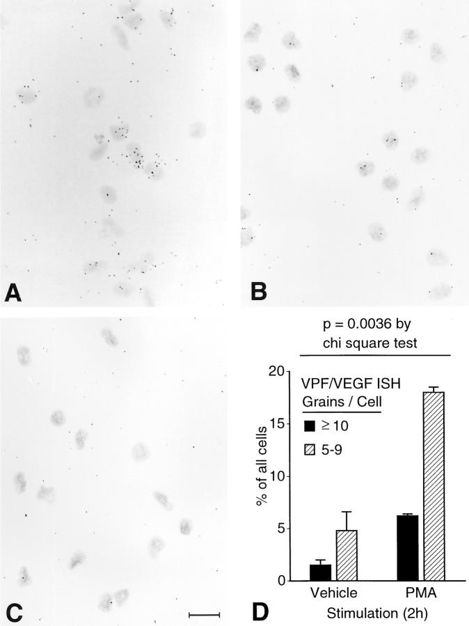
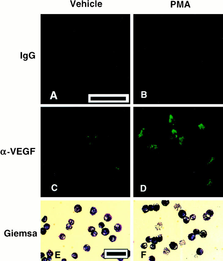
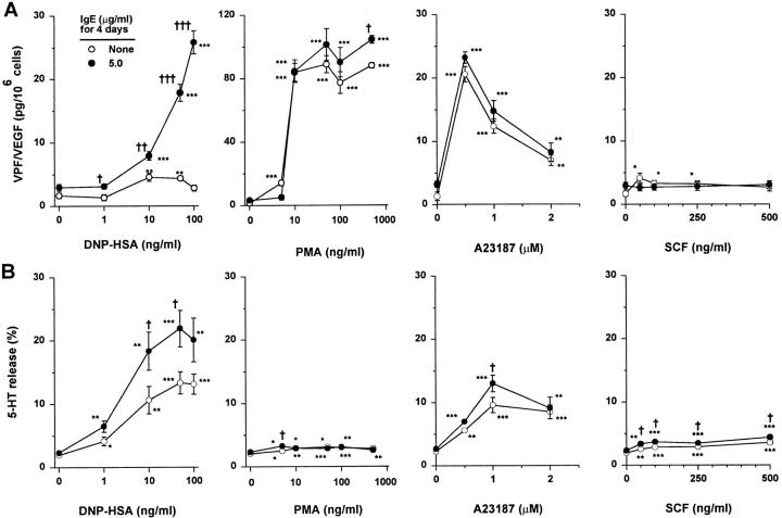
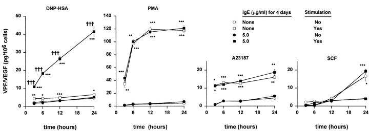
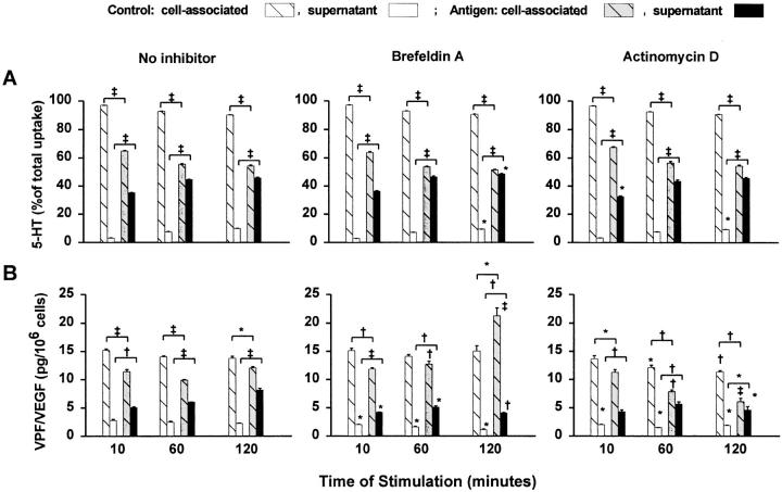
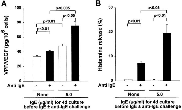
Vascular permeability factor/vascular endothelial cell growth factor (VPF/VEGF) can both potently enhance vascular permeability and induce proliferation of vascular endothelial cells. We report here that mouse or human mast cells can produce and secrete VPF/VEGF. Mouse mast cells release VPF/VEGF upon stimulation through Fcepsilon receptor I (FcepsilonRI) or c-kit, or after challenge with the protein kinase C activator, phorbol myristate acetate, or the calcium ionophore, A23187; such mast cells can rapidly release VPF/VEGF, apparently from a preformed pool, and can then sustain release by secreting newly synthesized protein. Notably, the Fc epsilonRI-dependent secretion of VPF/VEGF by either mouse or human mast cells can be significantly increased in cells which have undergone upregulation of Fc epsilonRI surface expression by a 4-d preincubation with immunoglobulin E. These findings establish that at least one cell type, the mast cell, can be stimulated to secrete VPF/VEGF upon immunologically specific activation via a member of the multichain immune recognition receptor family. Our observations also identify a new mechanism by which mast cells can contribute to enhanced vascular permeability and/or angiogenesis, in both allergic diseases and other settings.


Figures

Figure 1
Figure 1
Northern blot analysis of total RNA from C1.MC/C57.1 mast cells stimulated through FcεRI (A) or with PMA (B). C1.MC/ C57.1 cells were sensitized with IgE (10 μg IgE/ml) for 30 min, washed, and then stimulated with either 50 ng/ml DNP-HSA or 50 ng/ml PMA. Control cells (at 0.5 and 24 h) were challenged with medium (in A) or vehicle (0.005% DMSO, in B). Blotted total RNA from cells harvested at various times after stimulation was probed for VPF/VEGF and G3PDH (to demonstrate equal RNA loading).
Figure 2
Figure 2
ISH studies of freshly isolated mouse PMCs. Expression of VPF/VEGF mRNA is higher in mast cells that had been treated with PMA (50 ng/ml) for 2 h before hybridization to an 35S-labeled antisense riboprobe (A) than in vehicle-treated (control) mast cells that had been hybridized to the same antisense riboprobe (B). No specific signal is seen in PMA-stimulated mast cells hybridized to the 35S-labeled sense (control) riboprobe (C  ). Bar = 16 μm. (D) Graphic depiction of VPF/VEGF expression by PMA- versus vehicle-treated mast cells.
Figure 3
Figure 3
Immunofluorescent detection of VPF/VEGF immunoreactivity in a highly purified (99% pure) population of freshly isolated mouse PMCs. (A and B) Little or no staining of PMCs that had been incubated for 2 h with vehicle alone (A) or PMA (50 ng/ml; B) before incubation with a control rabbit IgG preparation (IgG). (C and D) VPF/ VEGF immunoreactivity was detected with a rabbit anti-VPF/ VEGF (α-VEGF  ) antibody in some PMCs that had been incubated for 2 h with either vehicle alone (C  ) or PMA (50 ng/ml; D). Black bar in A (for A–D) = 50 μm. (E and F  ) Giemsa-stained cytospin preparations of the same vehicle- (E  ) or PMA- (F  ) stimulated purified PMCs that were used for immunofluorescent detection of immunoreactivity (A–D). Black bar in E (for E and F  ) = 50 μm.
Figure 4
Figure 4
Release of (A) VPF/VEGF (in 6 h) and (B) serotonin (5-HT, in 10 min) from BMCMCs that exhibit different levels of surface expression of FcεRI, after challenge with various concentrations of either specific antigen (DNP-HSA), the protein kinase C activator PMA, the calcium ionophore A23187, or SCF. BALB/c BMCMCs were cultured without IgE or with IgE at 5.0 μg/ml for 4 d before passive sensitization and antigen challenge or challenge with A23187, PMA, or SCF for 6 h. 0 in the PMA and A23187 experiments refers to cells incubated without PMA or A23187, respectively, but in the highest concentration of vehicle used for these studies (DMSO at 0.5%). All values are mean ± SEM (n = 8/point) of data pooled from two experiments with different batches of BMCMCs (each n = 4/point) that gave very similar results. *P < 0.05, **P < 0.005, or ***P < 0.0001 versus corresponding control values for unstimulated or vehicle-treated cells. P < 0.05, †† P < 0.005, or ††† P < 0.0001 versus corresponding values for cells cultured without IgE for 4 d. Note the different VPF/VEGF scale for PMA stimulation.
Figure 5
Figure 5
Kinetics of the release of VPF/VEGF from BALB/c BMCMCs that exhibit different levels of surface expression of FcεRI, after challenge with either specific antigen (DNP-HSA, at 50 ng/ml), PMA (50 ng/ml), A23187 (1 μM), or SCF (500 ng/ml). BMCMCs were cultured without IgE or with IgE at 5.0 μg/ml for 4 d before passive sensitization and antigen challenge or challenge with A23187, PMA, or SCF. Cells used as unstimulated controls for DNP-HSA– or SCF-stimulated cells were maintained in medium alone. PMA- or A23187-stimulated cells were compared with cells maintained in medium containing vehicle (DMSO at 0.1%). All values are mean ± SEM (n = 8/point) of data pooled from two experiments with different batches of BMCMCs (each n = 4/point) that gave very similar results. *P < 0.05, **P < 0.005, or ***P < 0.0001 versus corresponding control values for unstimulated or vehicle-treated cells. P < 0.05, †† P < 0.005, or ††† P < 0.0001 versus corresponding values for cells cultured without IgE for 4 d. Note the different VPF/VEGF scale for PMA.
Figure 6
Figure 6
Kinetics of release of 5-HT (A) and VPF/VEGF (B) from BMCMCs stimulated through the FcεRI. Mast cells were sensitized with IgE (10 μg IgE/ml) for 2 h and then preincubated as indicated for 5 min at 37°C with either no inhibitor, the inhibitor of intracellular protein transport Brefeldin A (5 μg/ ml), or the transcription inhibitor Act D (10 μg/ml). Cells were then challenged for various time periods with DNP-HSA (Antigen) or medium (Control  ). Data for VPF/VEGF are mean ± SEM (n = 3/point); data for [3H]5-HT are mean ± SEM (n = 4/point). *P < 0.05, P < 0.005, P < 0.0001 versus values for the same time point in cells incubated without the inhibitor, or, as indicated (brackets), versus corresponding values for control cells that were not challenged with DNP-HSA. A second identical experiment with BMCMCs gave very similar results.
Figure 7
Figure 7
IgE-induced enhancement of FcεRI expression in human mast cells increases the ability of these cells to release VPF/VEGF (over a 16-h period; A) and histamine (over a 1-h period; B) in response to FcεRI cross-linking. HMCs (12 wk of culture, 85% mast cell purity) were cultured without or with human myeloma IgE at 5 μg/ml for 4 d before further passive sensitization with IgE and challenge with goat anti–human IgE antibody. The data shown (mean ± SEM; n = 3/bar) are representative of the results obtained in three separate experiments using mast cells derived from three different cord blood donors.

Similar articles

Cited by

References

    1. Selye, H. 1965. The Mast Cells. Butterworth Inc., Washington, DC. 488 pp.
    1. Galli SJ, Zsebo KM, Geissler EN. The kit ligand, stem cell factor. Adv Immunol. 1994;55:1–96. - PubMed
    1. Glowacki J, Mulliken JB. Mast cells in hemangiomas and vascular malformations. Pediatrics. 1982;70:48–51. - PubMed
    1. Lauria de Cidre L, Sacerdote de Lustig E. Mast cell kinetics during tumor growth. Tumor Biol. 1990;11:196–201. - PubMed
    1. Starkey JR, Crowle PK, Taubenberger S. Mast-cell-deficient W/Wvmice exhibit a decreased rate of tumor angiogenesis. Int J Cancer. 1988;42:48–52. - PubMed

Publication types

MeSH terms